Yuan Li, Yuhan Jiang, Bingbing Gao, Na Liu, Yukun Zhang, Huiling Zhou, Qingwei Song, Nan Wang, Yanwei Miao
{"title":"Regional high iron deposition on brain quantitative susceptibility mapping correlates with cognitive decline in chronic kidney disease patients.","authors":"Yuan Li, Yuhan Jiang, Bingbing Gao, Na Liu, Yukun Zhang, Huiling Zhou, Qingwei Song, Nan Wang, Yanwei Miao","doi":"10.1007/s11682-025-00976-0","DOIUrl":null,"url":null,"abstract":"<p><p>This study aimed to evaluate changes in gray matter nuclei iron deposition in chronic kidney disease (CKD) patients using the quantitative susceptibility mapping (QSM) threshold method, and analyze the relationship between brain iron levels and cognitive function. A total of fifty-three CKD patients were prospectively recruited, comprising 35 hemodialysis (HD, 57.54 ± 10.42 years, 21 males) and 18 non-hemodialysis (NHD, 55.06 ± 11.47 years, 10 males ), and were compared to 43 healthy controls (HC, 55.67 ± 7.79 years, 18 males). All participants underwent clinical assessments, neuropsychological tests, and QSM scans. The mean magnetic susceptibility value (MSV) and volume of the whole nuclei (MSV<sub>M</sub>, V<sub>M</sub>) and high iron region (MSV<sub>RII</sub>, V<sub>RII</sub>) were measured. Correlations between QSM data, neuropsychological scores, and clinical variables in HD group were analyzed. Linear regression analysis was performed to explore the effect of iron deposition on cognition and emotional well-being in HD group. A statistically significant P-value was set at 0.05. HD patients exhibited higher MSV<sub>M</sub> in the right red nucleus (RN) compared to HCs (P = 0.006). Additionally, significant differences in the MSV<sub>RII</sub> were observed in the left caudate nucleus (CN), bilateral putamen (Put), and right RN among the three groups (all P = 0.027, FDR-corrected). MSV<sub>RII</sub> of the left Put was positively correlated with creatinine and uric acid levels, while the MSV<sub>RII</sub> of the right Put was negatively correlated with mean corpuscular hemoglobin and mean corpuscular hemoglobin concentration. Regression analysis revealed that iron deposition in left CN was independently associated with depression, while iron deposition in left Put and right RN were independently positively associated with delayed recall performance. Conversely, iron deposition in bilateral Put and right RN were negatively associated with orientation ability, after controlling for age, sex, years of education and duration of dialysis. Brain iron deposition is often excessive and uneven in CKD patients, particularly those undergoing hemodialysis. Assessing regional high-iron deposition can provide valuable insights into the distribution of iron, which is associated with cognitive dysfunction and emotional disorders.</p>","PeriodicalId":9192,"journal":{"name":"Brain Imaging and Behavior","volume":" ","pages":"395-406"},"PeriodicalIF":2.4000,"publicationDate":"2025-04-01","publicationTypes":"Journal Article","fieldsOfStudy":null,"isOpenAccess":false,"openAccessPdf":"https://www.ncbi.nlm.nih.gov/pmc/articles/PMC11978685/pdf/","citationCount":"0","resultStr":null,"platform":"Semanticscholar","paperid":null,"PeriodicalName":"Brain Imaging and Behavior","FirstCategoryId":"3","ListUrlMain":"https://doi.org/10.1007/s11682-025-00976-0","RegionNum":3,"RegionCategory":"医学","ArticlePicture":[],"TitleCN":null,"AbstractTextCN":null,"PMCID":null,"EPubDate":"2025/2/11 0:00:00","PubModel":"Epub","JCR":"Q2","JCRName":"NEUROIMAGING","Score":null,"Total":0}
引用次数: 0
Abstract
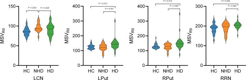
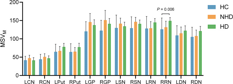
This study aimed to evaluate changes in gray matter nuclei iron deposition in chronic kidney disease (CKD) patients using the quantitative susceptibility mapping (QSM) threshold method, and analyze the relationship between brain iron levels and cognitive function. A total of fifty-three CKD patients were prospectively recruited, comprising 35 hemodialysis (HD, 57.54 ± 10.42 years, 21 males) and 18 non-hemodialysis (NHD, 55.06 ± 11.47 years, 10 males ), and were compared to 43 healthy controls (HC, 55.67 ± 7.79 years, 18 males). All participants underwent clinical assessments, neuropsychological tests, and QSM scans. The mean magnetic susceptibility value (MSV) and volume of the whole nuclei (MSVM, VM) and high iron region (MSVRII, VRII) were measured. Correlations between QSM data, neuropsychological scores, and clinical variables in HD group were analyzed. Linear regression analysis was performed to explore the effect of iron deposition on cognition and emotional well-being in HD group. A statistically significant P-value was set at 0.05. HD patients exhibited higher MSVM in the right red nucleus (RN) compared to HCs (P = 0.006). Additionally, significant differences in the MSVRII were observed in the left caudate nucleus (CN), bilateral putamen (Put), and right RN among the three groups (all P = 0.027, FDR-corrected). MSVRII of the left Put was positively correlated with creatinine and uric acid levels, while the MSVRII of the right Put was negatively correlated with mean corpuscular hemoglobin and mean corpuscular hemoglobin concentration. Regression analysis revealed that iron deposition in left CN was independently associated with depression, while iron deposition in left Put and right RN were independently positively associated with delayed recall performance. Conversely, iron deposition in bilateral Put and right RN were negatively associated with orientation ability, after controlling for age, sex, years of education and duration of dialysis. Brain iron deposition is often excessive and uneven in CKD patients, particularly those undergoing hemodialysis. Assessing regional high-iron deposition can provide valuable insights into the distribution of iron, which is associated with cognitive dysfunction and emotional disorders.
期刊介绍:
Brain Imaging and Behavior is a bi-monthly, peer-reviewed journal, that publishes clinically relevant research using neuroimaging approaches to enhance our understanding of disorders of higher brain function. The journal is targeted at clinicians and researchers in fields concerned with human brain-behavior relationships, such as neuropsychology, psychiatry, neurology, neurosurgery, rehabilitation, and cognitive neuroscience.

 求助内容:
求助内容: 应助结果提醒方式:


