Abbas Habibalahi, Ayad G Anwer, Aline Knab, Shane T Grey, Ewa M Goldys, Jared M Campbell
{"title":"Multispectral autofluorescence for label free classification of immune cell type and activation/polarization status.","authors":"Abbas Habibalahi, Ayad G Anwer, Aline Knab, Shane T Grey, Ewa M Goldys, Jared M Campbell","doi":"10.1111/sji.70004","DOIUrl":null,"url":null,"abstract":"<p><p>Evaluating immune status is a challenging and time-consuming process that involves analysing various biomarkers through numerous assays. The sensitive label-free technique of multispectral imaging of cell autofluorescence involves directly assessing the molecular composition of cells to gather biological information. Cells were cultured in RPMI 1640 modified media supplemented with penicillin-streptomycin and 10% foetal bovine serum at 37°C, with 5% CO<sub>2</sub> and 95% humidity. Activation and differentiation was confirmed using immunofluorophores against relevant markers. Multispectral microscopy utilized defined spectral regions, which spanned the excitation (345-476 nm) and emission (414-675 nm) wavelength ranges. In total, 56 distinct spectral channels were applied. These channels cover the spectrum of several fluorophores notably NAD(P)H and flavins, whose concentrations depend on cellular metabolism. We identified distinct spectral signatures for characterizing cells from the Jurkat, Ramos, THP-1, and HL-60 immune cell lines. These signatures correspond to four major immune cell types: T cells (Lymphocytes), B cells (Lymphocytes), monocytes and neutrophils. Moreover, our investigation explored the potential identification of both activated and resting forms of these cells, including the discrimination of M0, M1 and M2 polarized macrophages. Classification accuracy ranged from 92% to 100% based on receiver operator characteristic area under the curve (ROC AUC) assessment. These results indicate that the multispectral evaluation of cell autofluorescence is applicable for characterization of immune status. This includes the assessment of cell types and their activation status, all achievable through a single non-invasive assay.</p>","PeriodicalId":21493,"journal":{"name":"Scandinavian Journal of Immunology","volume":"101 2","pages":"e70004"},"PeriodicalIF":1.6000,"publicationDate":"2025-02-01","publicationTypes":"Journal Article","fieldsOfStudy":null,"isOpenAccess":false,"openAccessPdf":"https://www.ncbi.nlm.nih.gov/pmc/articles/PMC11808199/pdf/","citationCount":"0","resultStr":null,"platform":"Semanticscholar","paperid":null,"PeriodicalName":"Scandinavian Journal of Immunology","FirstCategoryId":"3","ListUrlMain":"https://doi.org/10.1111/sji.70004","RegionNum":4,"RegionCategory":"医学","ArticlePicture":[],"TitleCN":null,"AbstractTextCN":null,"PMCID":null,"EPubDate":"","PubModel":"","JCR":"Q2","JCRName":"IMMUNOLOGY","Score":null,"Total":0}
引用次数: 0
Abstract
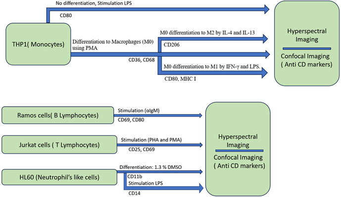
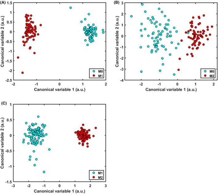
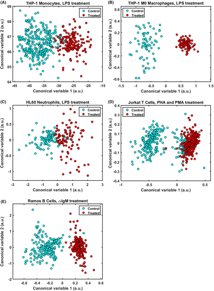
Evaluating immune status is a challenging and time-consuming process that involves analysing various biomarkers through numerous assays. The sensitive label-free technique of multispectral imaging of cell autofluorescence involves directly assessing the molecular composition of cells to gather biological information. Cells were cultured in RPMI 1640 modified media supplemented with penicillin-streptomycin and 10% foetal bovine serum at 37°C, with 5% CO2 and 95% humidity. Activation and differentiation was confirmed using immunofluorophores against relevant markers. Multispectral microscopy utilized defined spectral regions, which spanned the excitation (345-476 nm) and emission (414-675 nm) wavelength ranges. In total, 56 distinct spectral channels were applied. These channels cover the spectrum of several fluorophores notably NAD(P)H and flavins, whose concentrations depend on cellular metabolism. We identified distinct spectral signatures for characterizing cells from the Jurkat, Ramos, THP-1, and HL-60 immune cell lines. These signatures correspond to four major immune cell types: T cells (Lymphocytes), B cells (Lymphocytes), monocytes and neutrophils. Moreover, our investigation explored the potential identification of both activated and resting forms of these cells, including the discrimination of M0, M1 and M2 polarized macrophages. Classification accuracy ranged from 92% to 100% based on receiver operator characteristic area under the curve (ROC AUC) assessment. These results indicate that the multispectral evaluation of cell autofluorescence is applicable for characterization of immune status. This includes the assessment of cell types and their activation status, all achievable through a single non-invasive assay.
期刊介绍:
This peer-reviewed international journal publishes original articles and reviews on all aspects of basic, translational and clinical immunology. The journal aims to provide high quality service to authors, and high quality articles for readers.
The journal accepts for publication material from investigators all over the world, which makes a significant contribution to basic, translational and clinical immunology.
 求助内容:
求助内容: 应助结果提醒方式:


