Serge Szmukler-Moncler, David Morales Schwarz, Jorge Manuel Perez, Florian Beuer
{"title":"A digital way to assess the stain parameters that lead to soft tissue blanching when delivering an implant-supported crown.","authors":"Serge Szmukler-Moncler, David Morales Schwarz, Jorge Manuel Perez, Florian Beuer","doi":"10.1186/s40729-025-00598-7","DOIUrl":null,"url":null,"abstract":"<p><strong>Background: </strong>Dental implant systems provide standard cylindrical healing abutments of various diameters; however, they do not match the larger shape of the complex emergence profile of the prosthetic crowns. Adaptation of the soft tissues from a circular emergence profile to the one that suits the prosthetic crown involves a simultaneous squeezing and stretching of the gingiva. Often, this translates into local blanching of the gingiva and the prosthodontist must assess that blanching is transient. There is no literature about how much strain exerted by the prosthetic crown is leading or not to gingiva blanching. Aim of this paper is to present a digital workflow that allows measuring, upon prosthesis delivery, how much the strained gingiva is displaced under the crown and leads or not to blanching of the peri-implant soft tissues.</p><p><strong>Method and results: </strong>The digital workflow involves 3 intra-oral scans (IOS), IOS#1 at completion of the soft tissue healing, IOS#2 at prosthesis delivery, IOS#3 after soft tissue conditioning, and the STL files of the healing cap, the abutment, the implant and the prosthetic crown. The above are superposed and merged following a dedicated protocol that provides access to the distance the delivered crown deforms the strained gingiva. The present case study displayed distinct blanching intensities. Severe blanching was present when the strains applied to the gingiva caused a displacement of 1.3 mm and above; a displacement of 0.9 mm led to moderate blanching. No blanching was observed up to a displacement of 0.6 mm.</p><p><strong>Conclusion: </strong>A digital protocol, involving the superposition and merging of IOSs taken along a defined timeline and STLs of the implant hardware, allowed measuring the displacement distances a prosthetic crown wields upon delivery on the gingiva beneath the prosthesis. Various intensities of gingiva blanching could be related to distinct displacement distances of the healed gingiva that were triggered by attaching a prosthetic crown to the implant neck.</p>","PeriodicalId":14076,"journal":{"name":"International Journal of Implant Dentistry","volume":"11 1","pages":"10"},"PeriodicalIF":4.0000,"publicationDate":"2025-02-10","publicationTypes":"Journal Article","fieldsOfStudy":null,"isOpenAccess":false,"openAccessPdf":"https://www.ncbi.nlm.nih.gov/pmc/articles/PMC11811304/pdf/","citationCount":"0","resultStr":null,"platform":"Semanticscholar","paperid":null,"PeriodicalName":"International Journal of Implant Dentistry","FirstCategoryId":"3","ListUrlMain":"https://doi.org/10.1186/s40729-025-00598-7","RegionNum":3,"RegionCategory":"医学","ArticlePicture":[],"TitleCN":null,"AbstractTextCN":null,"PMCID":null,"EPubDate":"","PubModel":"","JCR":"Q1","JCRName":"DENTISTRY, ORAL SURGERY & MEDICINE","Score":null,"Total":0}
引用次数: 0
Abstract
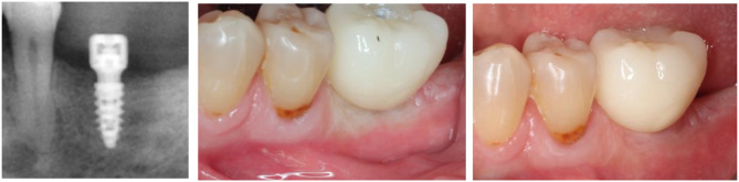
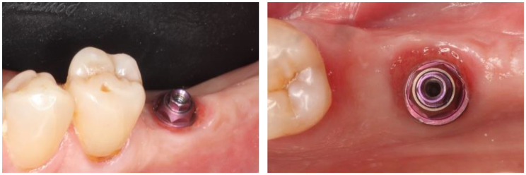
Background: Dental implant systems provide standard cylindrical healing abutments of various diameters; however, they do not match the larger shape of the complex emergence profile of the prosthetic crowns. Adaptation of the soft tissues from a circular emergence profile to the one that suits the prosthetic crown involves a simultaneous squeezing and stretching of the gingiva. Often, this translates into local blanching of the gingiva and the prosthodontist must assess that blanching is transient. There is no literature about how much strain exerted by the prosthetic crown is leading or not to gingiva blanching. Aim of this paper is to present a digital workflow that allows measuring, upon prosthesis delivery, how much the strained gingiva is displaced under the crown and leads or not to blanching of the peri-implant soft tissues.
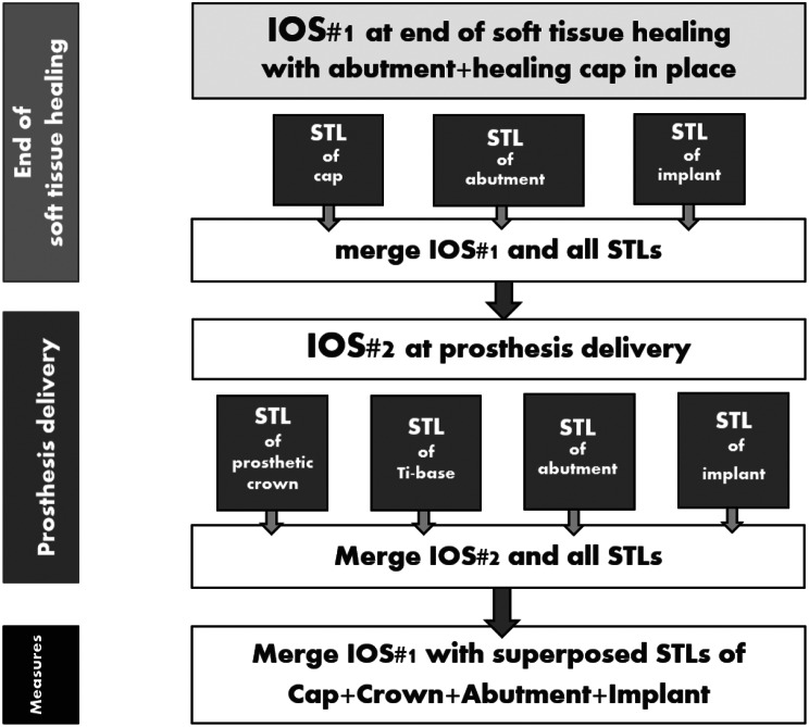
Method and results: The digital workflow involves 3 intra-oral scans (IOS), IOS#1 at completion of the soft tissue healing, IOS#2 at prosthesis delivery, IOS#3 after soft tissue conditioning, and the STL files of the healing cap, the abutment, the implant and the prosthetic crown. The above are superposed and merged following a dedicated protocol that provides access to the distance the delivered crown deforms the strained gingiva. The present case study displayed distinct blanching intensities. Severe blanching was present when the strains applied to the gingiva caused a displacement of 1.3 mm and above; a displacement of 0.9 mm led to moderate blanching. No blanching was observed up to a displacement of 0.6 mm.
Conclusion: A digital protocol, involving the superposition and merging of IOSs taken along a defined timeline and STLs of the implant hardware, allowed measuring the displacement distances a prosthetic crown wields upon delivery on the gingiva beneath the prosthesis. Various intensities of gingiva blanching could be related to distinct displacement distances of the healed gingiva that were triggered by attaching a prosthetic crown to the implant neck.
期刊介绍:
The International Journal of Implant Dentistry is a peer-reviewed open access journal published under the SpringerOpen brand. The journal is dedicated to promoting the exchange and discussion of all research areas relevant to implant dentistry in the form of systematic literature or invited reviews, prospective and retrospective clinical studies, clinical case reports, basic laboratory and animal research, and articles on material research and engineering.
 求助内容:
求助内容: 应助结果提醒方式:


