{"title":"Three-dimensional architecture of the palmar plate of the thumb metacarpophalangeal joint in infant macaque.","authors":"Hiroko Sato, Tatsuo Shimada, Tsuguaki Hosoyama, Yutaro Shibuta, Nobuhiro Kaku","doi":"10.1007/s00795-025-00423-5","DOIUrl":null,"url":null,"abstract":"<p><p>The palmar plate is a crucial structural part of hand, associated with metacarpophalangeal and interphalangeal joints. Pediatric disorders involving the palmar plate of thumb metacarpophalangeal joint include trigger thumb, hyperextension, instability, and dislocation. While anatomical differences exist between children and adults, detailed microstructure evaluations in infants remain unexplored. In this study, we provide a histological and structural assessment of the previously unresolved microstructure of the palmar plate in the thumb metacarpophalangeal joint of infant Japanese macaques (Cercopithecidae, Macaca fuscata), a relevant model for human development. Histological staining (light microscopy) and scanning electron microscopy were employed to visualize the three-dimensional microstructure. The palmar plate of the infant macaque was found to contain (1) elastic fibers, (2) hyaline cartilage composed of type II collagen, and (3) type I collagen fibers arranged in distinct patterns. The cartilaginous region exhibited a reticulate fiber arrangement on its periphery, while the membranous region displayed dense and complex fibers on the proximal phalanx side and parallel on the metacarpal side, respectively. This is the first comprehensive three-dimensional investigation of the infant's thumb's palmar microanatomy, providing a foundation for understanding its development and implications for pediatric disorders.</p>","PeriodicalId":18338,"journal":{"name":"Medical Molecular Morphology","volume":" ","pages":"163-170"},"PeriodicalIF":1.1000,"publicationDate":"2025-09-01","publicationTypes":"Journal Article","fieldsOfStudy":null,"isOpenAccess":false,"openAccessPdf":"https://www.ncbi.nlm.nih.gov/pmc/articles/PMC12378775/pdf/","citationCount":"0","resultStr":null,"platform":"Semanticscholar","paperid":null,"PeriodicalName":"Medical Molecular Morphology","FirstCategoryId":"3","ListUrlMain":"https://doi.org/10.1007/s00795-025-00423-5","RegionNum":4,"RegionCategory":"医学","ArticlePicture":[],"TitleCN":null,"AbstractTextCN":null,"PMCID":null,"EPubDate":"2025/2/8 0:00:00","PubModel":"Epub","JCR":"Q3","JCRName":"PATHOLOGY","Score":null,"Total":0}
引用次数: 0
Abstract
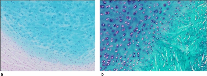
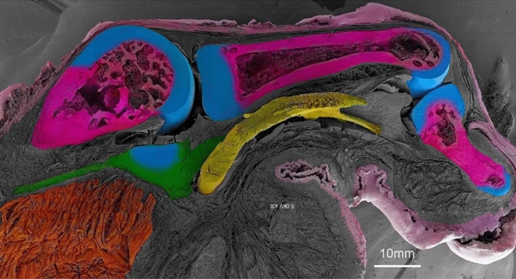
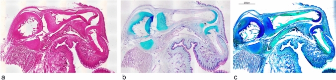
The palmar plate is a crucial structural part of hand, associated with metacarpophalangeal and interphalangeal joints. Pediatric disorders involving the palmar plate of thumb metacarpophalangeal joint include trigger thumb, hyperextension, instability, and dislocation. While anatomical differences exist between children and adults, detailed microstructure evaluations in infants remain unexplored. In this study, we provide a histological and structural assessment of the previously unresolved microstructure of the palmar plate in the thumb metacarpophalangeal joint of infant Japanese macaques (Cercopithecidae, Macaca fuscata), a relevant model for human development. Histological staining (light microscopy) and scanning electron microscopy were employed to visualize the three-dimensional microstructure. The palmar plate of the infant macaque was found to contain (1) elastic fibers, (2) hyaline cartilage composed of type II collagen, and (3) type I collagen fibers arranged in distinct patterns. The cartilaginous region exhibited a reticulate fiber arrangement on its periphery, while the membranous region displayed dense and complex fibers on the proximal phalanx side and parallel on the metacarpal side, respectively. This is the first comprehensive three-dimensional investigation of the infant's thumb's palmar microanatomy, providing a foundation for understanding its development and implications for pediatric disorders.
期刊介绍:
Medical Molecular Morphology is an international forum for researchers in both basic and clinical medicine to present and discuss new research on the structural mechanisms and the processes of health and disease at the molecular level. The structures of molecules, organelles, cells, tissues, and organs determine their normal function. Disease is thus best understood in terms of structural changes in these different levels of biological organization, especially in molecules and molecular interactions as well as the cellular localization of chemical components. Medical Molecular Morphology welcomes articles on basic or clinical research in the fields of cell biology, molecular biology, and medical, veterinary, and dental sciences using techniques for structural research such as electron microscopy, confocal laser scanning microscopy, enzyme histochemistry, immunohistochemistry, radioautography, X-ray microanalysis, and in situ hybridization.
Manuscripts submitted for publication must contain a statement to the effect that all human studies have been reviewed by the appropriate ethics committee and have therefore been performed in accordance with the ethical standards laid down in an appropriate version of the 1964 Declaration of Helsinki. It should also be stated clearly in the text that all persons gave their informed consent prior to their inclusion in the study. Details that might disclose the identity of the subjects under study should be omitted.
 求助内容:
求助内容: 应助结果提醒方式:


