Jie Su, Lantao Wang, Xiaoying Guan, Nan Li, Lixiao Sun
{"title":"Knocking-down annexin A3 suppresses inflammation, oxidative stress, apoptosis, and endoplasmic reticulum stress to attenuate sepsis-induced acute kidney injury in HK2 cells.","authors":"Jie Su, Lantao Wang, Xiaoying Guan, Nan Li, Lixiao Sun","doi":"10.25259/Cytojournal_64_2024","DOIUrl":null,"url":null,"abstract":"<p><strong>Objective: </strong>Sepsis-induced acute kidney injury (AKI) is considered as a life-threatening complication of sepsis. The purpose of this study is to clarify the involvement of annexin A3 (ANXA3) in sepsis-related AKI.</p><p><strong>Material and methods: </strong>Lipopolysaccharide (LPS) was used to establish a cell model based on HK2 cells. ANXA3 expression was quantified through quantitative real-time polymerase chain reaction. Cell proliferative capacities were assessed through 5-ethynyl-2'-deoxyuridine proliferation, cell counting kit-8, and colony formation experiments. Flow cytometry was utilized to analyze apoptotic cells. Inflammatory and oxidative stress indicators were measured by employing corresponding commercial assay kits. Endoplasmic reticulum (ER) stress markers were quantified through western blot analysis.</p><p><strong>Results: </strong>ANXA3 levels were significantly elevated in HK2 cells treated with LPS and in serum samples obtained from patients with AKI and sepsis (<i>P</i> < 0.001). LPS treatment exacerbated cellular damage, leading to increased ER and oxidative stresses, apoptosis, and inflammation, whereas knocking down ANXA3 significantly reversed these changes (<i>P</i> < 0.001).</p><p><strong>Conclusion: </strong>Interference with ANXA3 protected HK2 cells from LPS-induced cell injury through inhibiting inflammation, oxidative stress, apoptosis, and ER stress.</p>","PeriodicalId":49082,"journal":{"name":"Cytojournal","volume":"21 ","pages":"75"},"PeriodicalIF":3.1000,"publicationDate":"2024-12-27","publicationTypes":"Journal Article","fieldsOfStudy":null,"isOpenAccess":false,"openAccessPdf":"https://www.ncbi.nlm.nih.gov/pmc/articles/PMC11801658/pdf/","citationCount":"0","resultStr":null,"platform":"Semanticscholar","paperid":null,"PeriodicalName":"Cytojournal","FirstCategoryId":"3","ListUrlMain":"https://doi.org/10.25259/Cytojournal_64_2024","RegionNum":4,"RegionCategory":"医学","ArticlePicture":[],"TitleCN":null,"AbstractTextCN":null,"PMCID":null,"EPubDate":"2024/1/1 0:00:00","PubModel":"eCollection","JCR":"Q2","JCRName":"PATHOLOGY","Score":null,"Total":0}
引用次数: 0
Abstract
Objective: Sepsis-induced acute kidney injury (AKI) is considered as a life-threatening complication of sepsis. The purpose of this study is to clarify the involvement of annexin A3 (ANXA3) in sepsis-related AKI.
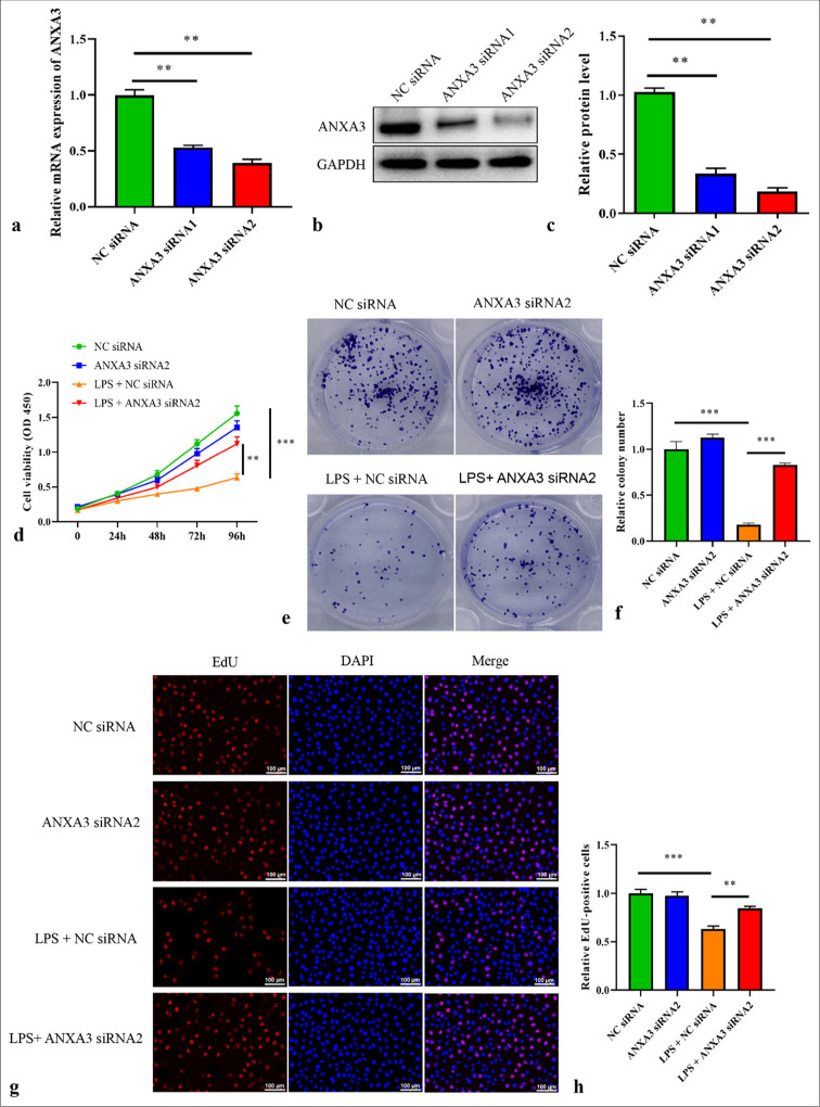
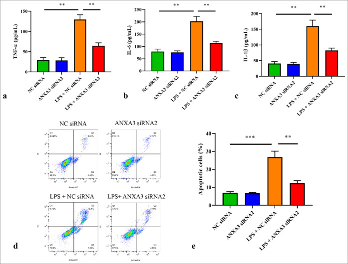
Material and methods: Lipopolysaccharide (LPS) was used to establish a cell model based on HK2 cells. ANXA3 expression was quantified through quantitative real-time polymerase chain reaction. Cell proliferative capacities were assessed through 5-ethynyl-2'-deoxyuridine proliferation, cell counting kit-8, and colony formation experiments. Flow cytometry was utilized to analyze apoptotic cells. Inflammatory and oxidative stress indicators were measured by employing corresponding commercial assay kits. Endoplasmic reticulum (ER) stress markers were quantified through western blot analysis.
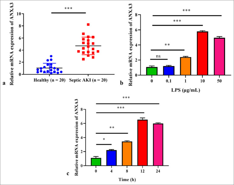
Results: ANXA3 levels were significantly elevated in HK2 cells treated with LPS and in serum samples obtained from patients with AKI and sepsis (P < 0.001). LPS treatment exacerbated cellular damage, leading to increased ER and oxidative stresses, apoptosis, and inflammation, whereas knocking down ANXA3 significantly reversed these changes (P < 0.001).
Conclusion: Interference with ANXA3 protected HK2 cells from LPS-induced cell injury through inhibiting inflammation, oxidative stress, apoptosis, and ER stress.
期刊介绍:
The CytoJournal is an open-access peer-reviewed journal committed to publishing high-quality articles in the field of Diagnostic Cytopathology including Molecular aspects. The journal is owned by the Cytopathology Foundation and published by the Scientific Scholar.
 求助内容:
求助内容: 应助结果提醒方式:


