Unexpected Detection of Cephalad Renal Ectopia Due to Large Omphalocele Containing the Liver on Tc-99m DMSA Scintigraphy.
IF 1.1
Q4 RADIOLOGY, NUCLEAR MEDICINE & MEDICAL IMAGING
Molecular Imaging and Radionuclide Therapy
Pub Date : 2025-02-07
DOI:10.4274/mirt.galenos.2024.68095
引用次数: 0
Abstract
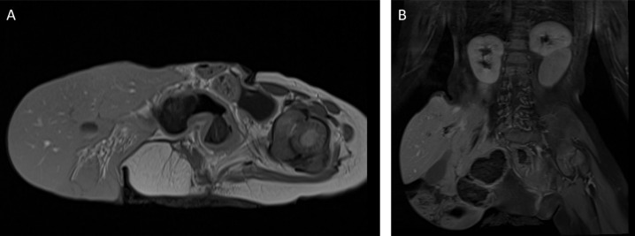
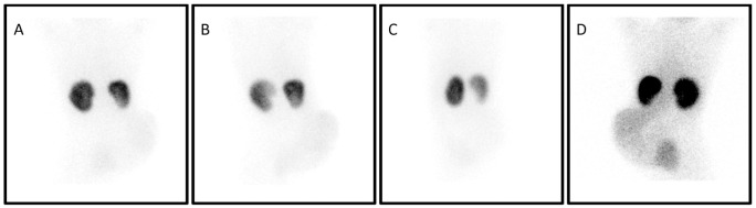
Omphalocele is a congenital abdominal wall defect with herniation of abdominal viscera into a sac. Tc-99m DMSA renal cortical scan is a functional imaging technique used for detecting parenchymal defects, mostly in patients with recurrent urinary tract infection as well as congenital renal abnormalities. Renal anomalies are known to accompany omphalocele. In this retrospective study, we present a case of cephalad renal ectopia as observed on Tc-99m DMSA scintigraphy in a patient with omphalocele due to a large hernia sac containing most of the liver; and we review the renal abnormalities associated with omphalocele in the literature.
Tc-99m DMSA显像意外发现含肝大脐膨出所致头肾异位。
脐膨出是一种先天性腹壁缺损,腹部脏器疝入囊内。Tc-99m DMSA肾皮质扫描是一种用于发现实质缺陷的功能成像技术,多用于复发性尿路感染和先天性肾脏异常患者。肾异常常伴有脐膨出。在这项回顾性研究中,我们报告了一例在Tc-99m DMSA显像上观察到的头肾异位患者,该患者因大疝囊包含大部分肝脏而导致脐膨出;我们回顾了文献中与脐膨出相关的肾脏异常。
本文章由计算机程序翻译,如有差异,请以英文原文为准。
求助全文
约1分钟内获得全文
求助全文
来源期刊

Molecular Imaging and Radionuclide Therapy
RADIOLOGY, NUCLEAR MEDICINE & MEDICAL IMAGING-
CiteScore
1.30
自引率
0.00%
发文量
50
期刊介绍:
Molecular Imaging and Radionuclide Therapy (Mol Imaging Radionucl Ther, MIRT) is publishes original research articles, invited reviews, editorials, short communications, letters, consensus statements, guidelines and case reports with a literature review on the topic, in the field of molecular imaging, multimodality imaging, nuclear medicine, radionuclide therapy, radiopharmacy, medical physics, dosimetry and radiobiology.
 求助内容:
求助内容: 应助结果提醒方式:


