{"title":"Image Analysis as tool for Predicting Colorectal Cancer Molecular Alterations: A Scoping Review.","authors":"Saman Mohammadpour, Hassan Emami, Reza Rabiei, Azamossadat Hosseini, Hamid Moghaddasi, Fariborz Faeghi, Rafat Bagherzadeh","doi":"10.4274/mirt.galenos.2024.86402","DOIUrl":null,"url":null,"abstract":"<p><strong>Objectives: </strong>Among the most important diagnostic indicators of colorectal cancer; however, measuring molecular alterations are invasive and expensive. This study aimed to investigate the application of image processing to predict molecular alterations in colorectal cancer.</p><p><strong>Methods: </strong>In this scoping review, we searched for relevant literature by searching the Web of Science, Scopus, and PubMed databases. The method of selecting the articles and reporting the findings was according to the guidelines of the Preferred Reporting Items for Systematic Reviews and Meta-Analyses; moreover, the Strengthening the Reporting of Observational Studies in Epidemiology checklist was used to assess the quality of the studies.</p><p><strong>Results: </strong>Sixty seven out of 2,223 articles, 67 were relevant to the aim of the study, and finally 41 studies with sufficient quality were reviewed. The prediction of Kirsten Rat Sarcoma Viral Oncogene Homolog (KRAS), Neuroblastoma RAS Viral (NRAS), B-Raf proto-oncogene, serine/threonine kinase (BRAF), Tumor Protein 53 (TP53), Adenomatous Polyposis Coli, and microsatellite instability (MSI) with the help of image analysis has received more attention than other molecular characteristics. The studies used computed tomography (CT), magnetic resonance imaging (MRI), and <sup>18</sup>F-FDG positron emission tomography (PET)/CT with radionics and quantitative analysis to predict molecular alterations in colorectal cancer, analyzing features like texture, maximum standard uptake value, and MTV using various statistical methods. In 39 studies, there was a significant relationship between the features extracted from these images and molecular alterations. Different modalities were used to measure the area under the receiver operating characteristic curve for predicting the alterations in KRAS, MSI, BRAF, and TP53, with an average of 78, 81, 80 and 71%, respectively.</p><p><strong>Conclusion: </strong>This scoping review underscores the potential of radiogenomics in predicting molecular alterations in colorectal cancer through non-invasive imaging modalities, like CT, MRI, and <sup>18</sup>F-FDG PET/CT. The analysis of 41 studies showed the appropriate prediction of key alterations, such as KRAS, NRAS, BRAF, TP53, and MSI, highlighting the promise of radionics and texture features in enhancing predictive accuracy.</p>","PeriodicalId":44681,"journal":{"name":"Molecular Imaging and Radionuclide Therapy","volume":"34 1","pages":"10-25"},"PeriodicalIF":1.1000,"publicationDate":"2025-02-07","publicationTypes":"Journal Article","fieldsOfStudy":null,"isOpenAccess":false,"openAccessPdf":"https://www.ncbi.nlm.nih.gov/pmc/articles/PMC11827529/pdf/","citationCount":"0","resultStr":null,"platform":"Semanticscholar","paperid":null,"PeriodicalName":"Molecular Imaging and Radionuclide Therapy","FirstCategoryId":"1085","ListUrlMain":"https://doi.org/10.4274/mirt.galenos.2024.86402","RegionNum":0,"RegionCategory":null,"ArticlePicture":[],"TitleCN":null,"AbstractTextCN":null,"PMCID":null,"EPubDate":"","PubModel":"","JCR":"Q4","JCRName":"RADIOLOGY, NUCLEAR MEDICINE & MEDICAL IMAGING","Score":null,"Total":0}
引用次数: 0
Abstract
Objectives: Among the most important diagnostic indicators of colorectal cancer; however, measuring molecular alterations are invasive and expensive. This study aimed to investigate the application of image processing to predict molecular alterations in colorectal cancer.
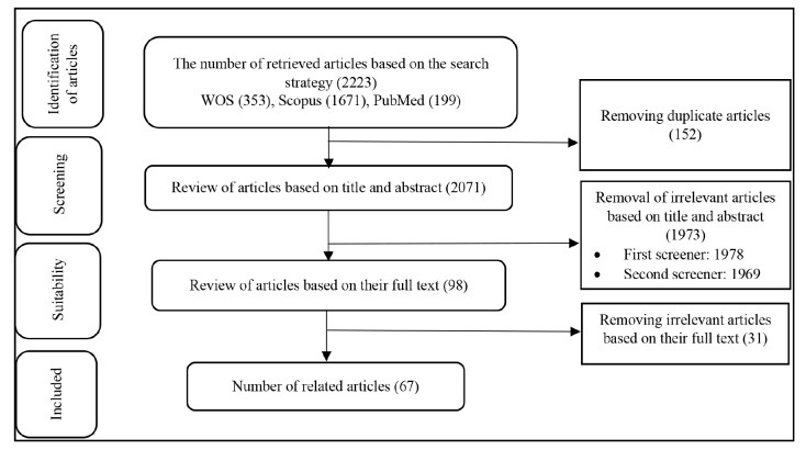
Methods: In this scoping review, we searched for relevant literature by searching the Web of Science, Scopus, and PubMed databases. The method of selecting the articles and reporting the findings was according to the guidelines of the Preferred Reporting Items for Systematic Reviews and Meta-Analyses; moreover, the Strengthening the Reporting of Observational Studies in Epidemiology checklist was used to assess the quality of the studies.
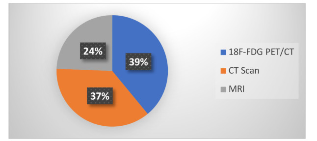
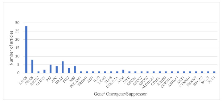
Results: Sixty seven out of 2,223 articles, 67 were relevant to the aim of the study, and finally 41 studies with sufficient quality were reviewed. The prediction of Kirsten Rat Sarcoma Viral Oncogene Homolog (KRAS), Neuroblastoma RAS Viral (NRAS), B-Raf proto-oncogene, serine/threonine kinase (BRAF), Tumor Protein 53 (TP53), Adenomatous Polyposis Coli, and microsatellite instability (MSI) with the help of image analysis has received more attention than other molecular characteristics. The studies used computed tomography (CT), magnetic resonance imaging (MRI), and 18F-FDG positron emission tomography (PET)/CT with radionics and quantitative analysis to predict molecular alterations in colorectal cancer, analyzing features like texture, maximum standard uptake value, and MTV using various statistical methods. In 39 studies, there was a significant relationship between the features extracted from these images and molecular alterations. Different modalities were used to measure the area under the receiver operating characteristic curve for predicting the alterations in KRAS, MSI, BRAF, and TP53, with an average of 78, 81, 80 and 71%, respectively.
Conclusion: This scoping review underscores the potential of radiogenomics in predicting molecular alterations in colorectal cancer through non-invasive imaging modalities, like CT, MRI, and 18F-FDG PET/CT. The analysis of 41 studies showed the appropriate prediction of key alterations, such as KRAS, NRAS, BRAF, TP53, and MSI, highlighting the promise of radionics and texture features in enhancing predictive accuracy.
期刊介绍:
Molecular Imaging and Radionuclide Therapy (Mol Imaging Radionucl Ther, MIRT) is publishes original research articles, invited reviews, editorials, short communications, letters, consensus statements, guidelines and case reports with a literature review on the topic, in the field of molecular imaging, multimodality imaging, nuclear medicine, radionuclide therapy, radiopharmacy, medical physics, dosimetry and radiobiology.

 求助内容:
求助内容: 应助结果提醒方式:


