Ahmet Tanyeri, Rıdvan Akbulut, Emir Hüseyin Nevai, Yakup Yürekli
{"title":"Correlation of 3T Diffusion-weighted MRI and <sup>18</sup>F-FDG-PET/CT in Liver Metastases: SUV Versus ADC.","authors":"Ahmet Tanyeri, Rıdvan Akbulut, Emir Hüseyin Nevai, Yakup Yürekli","doi":"10.4274/mirt.galenos.2024.37431","DOIUrl":null,"url":null,"abstract":"<p><strong>Objectives: </strong>Positron emission tomography/computed tomography (PET/CT) and magnetic resonance imaging (MRI) are widely used in the diagnosis and follow-up of liver metastases. Both modalities provide anatomical and functional information and have advantages and disadvantages. The objective of this study was to investigate the correlation between apparent diffusion coefficient (ADC) and standardized uptake value (SUV) values in metastatic liver lesions.</p><p><strong>Methods: </strong>Abdominal magnetic resonance (MR) scans performed between April 2021 and 2024 using the 3T MR scanner were retrospectively evaluated. Thirty-three patients with liver metastases, less than one month between magnetic resonance imaging (MRI) and PET/CT, no treatment during this period, and lesions larger than 1 cm were included in the study. In each MRI scan, an index lesion was selected for ADC measurement. The radiologist and nuclear medicine specialist measured the same index lesion without the patient being informed of the results.</p><p><strong>Results: </strong>The mean age of the 33 patients was 59±12 years, with 17 (51%) men and 16 (49%) women. The mean size of the index lesions was 27±9 mm. In MRI, mean ADC<sub>min</sub>: (0.54±0.2) ×10<sup>-3</sup>mm<sup>2</sup>/s; ADC<sub>mean</sub>: (1.02±0.2) ×10<sup>-3</sup>mm<sup>2</sup>/s; ADC<sub>max</sub>: (1.48±0.44) ×10<sup>-3</sup>mm<sup>2</sup>/s; and region of interest area was calculated as 6±4.6 cm<sup>2</sup>. In PET/CT, mean SUV<sub>mean</sub>: 5.8±3.3; SUV<sub>peak</sub>: 6.8±4.3; SUV<sub>max</sub>: 10.7±5.6; and metabolic tumor volume: 12.1 (7.4-20.7) cm<sup>3</sup>. No statistically significant correlation was found between ADC and SUV values.</p><p><strong>Conclusion: </strong>There was no correlation between ADC and SUV values in liver metastases. Prospective studies with a large patient group are needed.</p>","PeriodicalId":44681,"journal":{"name":"Molecular Imaging and Radionuclide Therapy","volume":"34 1","pages":"48-54"},"PeriodicalIF":1.1000,"publicationDate":"2025-02-07","publicationTypes":"Journal Article","fieldsOfStudy":null,"isOpenAccess":false,"openAccessPdf":"https://www.ncbi.nlm.nih.gov/pmc/articles/PMC11827527/pdf/","citationCount":"0","resultStr":null,"platform":"Semanticscholar","paperid":null,"PeriodicalName":"Molecular Imaging and Radionuclide Therapy","FirstCategoryId":"1085","ListUrlMain":"https://doi.org/10.4274/mirt.galenos.2024.37431","RegionNum":0,"RegionCategory":null,"ArticlePicture":[],"TitleCN":null,"AbstractTextCN":null,"PMCID":null,"EPubDate":"","PubModel":"","JCR":"Q4","JCRName":"RADIOLOGY, NUCLEAR MEDICINE & MEDICAL IMAGING","Score":null,"Total":0}
引用次数: 0
Abstract
Objectives: Positron emission tomography/computed tomography (PET/CT) and magnetic resonance imaging (MRI) are widely used in the diagnosis and follow-up of liver metastases. Both modalities provide anatomical and functional information and have advantages and disadvantages. The objective of this study was to investigate the correlation between apparent diffusion coefficient (ADC) and standardized uptake value (SUV) values in metastatic liver lesions.
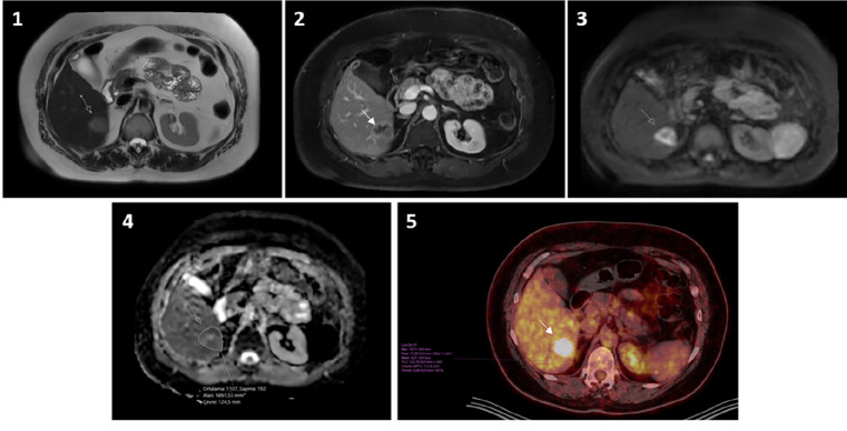
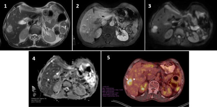
Methods: Abdominal magnetic resonance (MR) scans performed between April 2021 and 2024 using the 3T MR scanner were retrospectively evaluated. Thirty-three patients with liver metastases, less than one month between magnetic resonance imaging (MRI) and PET/CT, no treatment during this period, and lesions larger than 1 cm were included in the study. In each MRI scan, an index lesion was selected for ADC measurement. The radiologist and nuclear medicine specialist measured the same index lesion without the patient being informed of the results.
Results: The mean age of the 33 patients was 59±12 years, with 17 (51%) men and 16 (49%) women. The mean size of the index lesions was 27±9 mm. In MRI, mean ADCmin: (0.54±0.2) ×10-3mm2/s; ADCmean: (1.02±0.2) ×10-3mm2/s; ADCmax: (1.48±0.44) ×10-3mm2/s; and region of interest area was calculated as 6±4.6 cm2. In PET/CT, mean SUVmean: 5.8±3.3; SUVpeak: 6.8±4.3; SUVmax: 10.7±5.6; and metabolic tumor volume: 12.1 (7.4-20.7) cm3. No statistically significant correlation was found between ADC and SUV values.
Conclusion: There was no correlation between ADC and SUV values in liver metastases. Prospective studies with a large patient group are needed.
期刊介绍:
Molecular Imaging and Radionuclide Therapy (Mol Imaging Radionucl Ther, MIRT) is publishes original research articles, invited reviews, editorials, short communications, letters, consensus statements, guidelines and case reports with a literature review on the topic, in the field of molecular imaging, multimodality imaging, nuclear medicine, radionuclide therapy, radiopharmacy, medical physics, dosimetry and radiobiology.

 求助内容:
求助内容: 应助结果提醒方式:


