{"title":"Saccade Dynamics in the Acute and Recovery Phase of Abducens Nerve Palsy.","authors":"Elissavet Kemanetzoglou, Klio Chatzistefanou, Nikolaos Smyrnis, Evangelia Kararizou, Evangelos Anagnostou","doi":"10.18502/jovr.v19i4.14429","DOIUrl":null,"url":null,"abstract":"<p><strong>Purpose: </strong>To examine the natural adaptive course of ocular motor system in unilateral abducens nerve palsy while addressing the scarce literature on saccade dynamics and natural adaptation.</p><p><strong>Methods: </strong>Binocular horizontal eye movements were recorded from 18 healthy adults and 21 adults with unilateral abducens nerve palsy during the acute and chronic phases. Dynamics of the paretic and non-paretic eyes were compared, and the non-paretic eye dynamics were correlated with the respective prism diopters. Non-parametric tests were used for statistical comparisons.</p><p><strong>Results: </strong>The paretic eye, compared to the non-paretic eye, presented a slightly lower saccadic gain and velocity/amplitude ratio and a higher duration/amplitude ratio. The non-paretic eye, compared to healthy controls, showed consistent amplitude gain ( <math><mo>></mo></math> 1) and a tendency for a higher duration/amplitude ratio. In the acute phase, when the non-paretic eye was covered, the paretic eye's amplitude ratio was lower and the duration/amplitude ratio decreased significantly. In the acute phase, a greater degree of esotropia in the paretic eye was associated with a lower amplitude gain and duration/amplitude ratio in the non-paretic eye.</p><p><strong>Conclusion: </strong>During adaptation in abducens nerve palsy, the saccade duration of the paretic eye increased, and a similar tendency was observed in the non-paretic eye. This finding likely reflects a change in the \"pulse-step\" pattern and may be related to plastic changes in central structures, such as the cerebellum, that support learning processes.</p>","PeriodicalId":16586,"journal":{"name":"Journal of Ophthalmic & Vision Research","volume":"19 4","pages":"449-458"},"PeriodicalIF":1.5000,"publicationDate":"2024-12-31","publicationTypes":"Journal Article","fieldsOfStudy":null,"isOpenAccess":false,"openAccessPdf":"https://www.ncbi.nlm.nih.gov/pmc/articles/PMC11795006/pdf/","citationCount":"0","resultStr":null,"platform":"Semanticscholar","paperid":null,"PeriodicalName":"Journal of Ophthalmic & Vision Research","FirstCategoryId":"1085","ListUrlMain":"https://doi.org/10.18502/jovr.v19i4.14429","RegionNum":0,"RegionCategory":null,"ArticlePicture":[],"TitleCN":null,"AbstractTextCN":null,"PMCID":null,"EPubDate":"2024/12/1 0:00:00","PubModel":"eCollection","JCR":"Q3","JCRName":"OPHTHALMOLOGY","Score":null,"Total":0}
引用次数: 0
Abstract
Purpose: To examine the natural adaptive course of ocular motor system in unilateral abducens nerve palsy while addressing the scarce literature on saccade dynamics and natural adaptation.
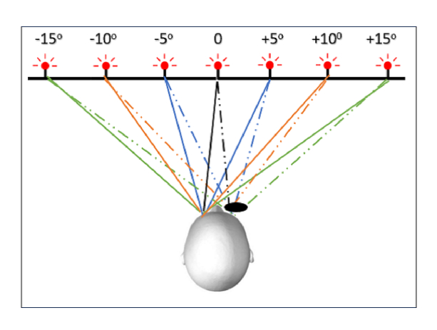
Methods: Binocular horizontal eye movements were recorded from 18 healthy adults and 21 adults with unilateral abducens nerve palsy during the acute and chronic phases. Dynamics of the paretic and non-paretic eyes were compared, and the non-paretic eye dynamics were correlated with the respective prism diopters. Non-parametric tests were used for statistical comparisons.
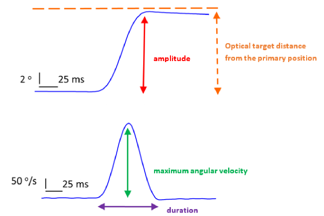
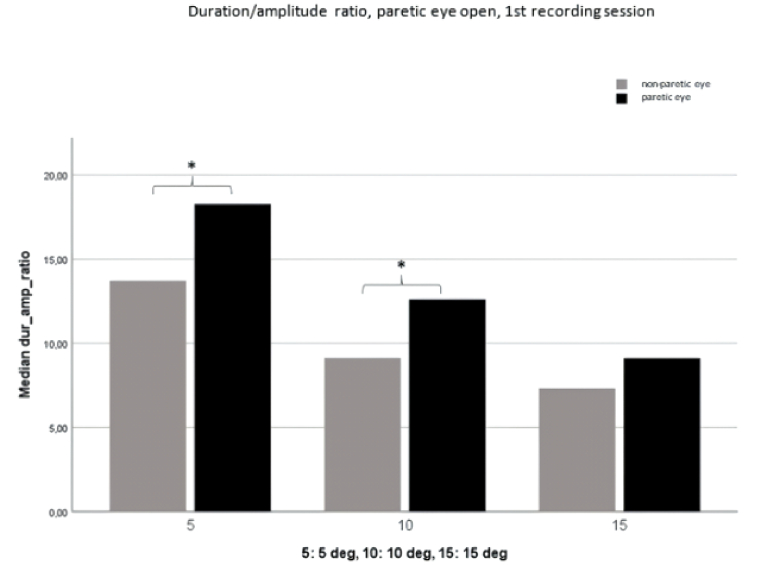
Results: The paretic eye, compared to the non-paretic eye, presented a slightly lower saccadic gain and velocity/amplitude ratio and a higher duration/amplitude ratio. The non-paretic eye, compared to healthy controls, showed consistent amplitude gain ( 1) and a tendency for a higher duration/amplitude ratio. In the acute phase, when the non-paretic eye was covered, the paretic eye's amplitude ratio was lower and the duration/amplitude ratio decreased significantly. In the acute phase, a greater degree of esotropia in the paretic eye was associated with a lower amplitude gain and duration/amplitude ratio in the non-paretic eye.
Conclusion: During adaptation in abducens nerve palsy, the saccade duration of the paretic eye increased, and a similar tendency was observed in the non-paretic eye. This finding likely reflects a change in the "pulse-step" pattern and may be related to plastic changes in central structures, such as the cerebellum, that support learning processes.

 求助内容:
求助内容: 应助结果提醒方式:


