Innovative MXene/SilMA-Based Conductive Bioink for Three Dimensional Bioprinting of Neural Stem Cell Spheroids in Neural Tissue Engineering
IF 8.2
2区 材料科学
Q1 MATERIALS SCIENCE, MULTIDISCIPLINARY
引用次数: 0
Abstract
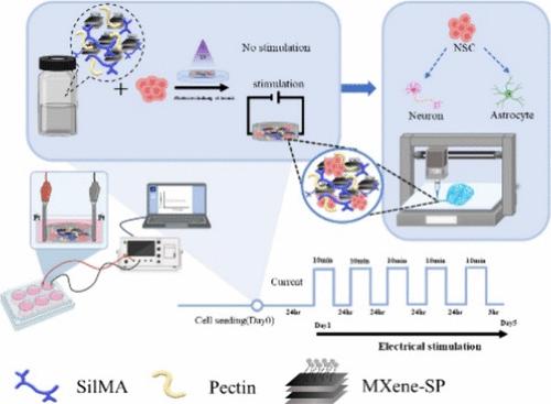
Conductive bioinks, integrated with 3D bioprinting and electrical stimulation, are essential for advancing neural tissue engineering. This study developed a SilMA/Pectin/MXene-soybean phospholipids (SP) bioink, where SilMA (silk fibroin modified with glycidyl methacrylate) provides a structural base, pectin enhances printability and shear-thinning properties, and MXene-SP improves conductivity through superior dispersibility. Increasing pectin and MXene-SP concentrations reduced the hydrogel’s Young’s modulus, promoting neural stem cell (NSC) differentiation into neurons. Electrochemical analyses revealed that higher MXene-SP levels decreased impedance and increased redox current, while conductivity measurements showed improved performance compared to unmodified MXene. NSCs encapsulated in the bioink achieved maximum proliferation under electrical stimulation at 300 μA for 10 min daily over 5 days. Neuronal differentiation positively correlated with MXene-SP concentration and stimulation intensity. Synaptic activity and vesicle recycling, assessed using FM1–43 dye, were significantly enhanced under electrical stimulation. This study successfully developed a biocompatible conductive bioink capable of inducing neuronal differentiation. Electrical stimulation further promoted cell proliferation, neuronal differentiation, and enhanced synaptic function. This bioink shows great potential for future applications in neural tissue engineering.
基于MXene/ silma的新型导电生物链用于神经组织工程中神经干细胞球体的三维生物打印
导电性生物墨水与3D生物打印和电刺激相结合,对推进神经组织工程至关重要。本研究开发了一种SilMA/Pectin/MXene-soybean磷脂(SP)生物链,其中SilMA(丝素蛋白改性与甲基丙烯酸缩水甘油酯)提供了结构基础,果胶提高了印刷性和剪切薄化性能,MXene-SP通过优异的分散性提高了导电性。增加果胶和MXene-SP浓度降低水凝胶的杨氏模量,促进神经干细胞(NSC)向神经元的分化。电化学分析表明,较高的MXene- sp水平降低了阻抗,增加了氧化还原电流,而电导率测量显示,与未改性的MXene相比,性能有所提高。在300 μA的电刺激下,每天10分钟,连续5天,包封在生物链中的NSCs增殖达到最大。神经元分化与MXene-SP浓度和刺激强度呈正相关。用FM1-43染料评估,突触活性和囊泡循环在电刺激下显著增强。本研究成功开发了一种具有诱导神经元分化能力的生物相容性导电生物链接。电刺激进一步促进细胞增殖、神经元分化,增强突触功能。这种生物链接在神经组织工程中显示出巨大的应用潜力。
本文章由计算机程序翻译,如有差异,请以英文原文为准。
求助全文
约1分钟内获得全文
求助全文
来源期刊
ACS Applied Materials & Interfaces
工程技术-材料科学:综合
CiteScore
16.00
自引率
6.30%
发文量
4978
审稿时长
1.8 months
期刊介绍:
ACS Applied Materials & Interfaces is a leading interdisciplinary journal that brings together chemists, engineers, physicists, and biologists to explore the development and utilization of newly-discovered materials and interfacial processes for specific applications. Our journal has experienced remarkable growth since its establishment in 2009, both in terms of the number of articles published and the impact of the research showcased. We are proud to foster a truly global community, with the majority of published articles originating from outside the United States, reflecting the rapid growth of applied research worldwide.
 求助内容:
求助内容: 应助结果提醒方式:


