Jiajia Tang, Jie Zhang, Yang Li, Yongzhi Hu, Doudou He, Hao Ni, Jiulou Zhang, Feiyun Wu, Yuxia Tang, Shouju Wang
{"title":"Interpretable Radiomics Model Predicts Nanomedicine Tumor Accumulation Using Routine Medical Imaging","authors":"Jiajia Tang, Jie Zhang, Yang Li, Yongzhi Hu, Doudou He, Hao Ni, Jiulou Zhang, Feiyun Wu, Yuxia Tang, Shouju Wang","doi":"10.1002/adma.202416696","DOIUrl":null,"url":null,"abstract":"<p>Accurately predicting nanomedicine accumulation is critical for guiding patient stratification and optimizing treatment strategies in the context of precision medicine. However, non-invasive prediction of nanomedicine accumulation remains challenging, primarily due to the complexity of identifying relevant imaging features that predict accumulation. Here, a novel non-invasive method is proposed that utilizes standard-of-care medical imaging modalities, including computed tomography and ultrasound, combined with a radiomics-based model to predict nanomedicine accumulation in tumor. The model is validated using a test dataset consisting of seven tumor xenografts in mice and three sizes of gold nanoparticles, achieving an area under the receiver operating characteristic curve of 0.851. The median accumulation levels of tumors predicted as “high accumulators” are 2.69 times greater than those predicted as “low accumulators”. Analysis of this machine-learning-driven interpretable radiomics model revealed imaging features that are strongly correlated with dense stroma, a recognized biological barrier to effective nanomedicine delivery. Radiomics-based prediction of tumor accumulation holds promise for stratifying patient and enabling precise tailoring of nanomedicine treatment strategies.</p>","PeriodicalId":114,"journal":{"name":"Advanced Materials","volume":"37 12","pages":""},"PeriodicalIF":26.8000,"publicationDate":"2025-02-07","publicationTypes":"Journal Article","fieldsOfStudy":null,"isOpenAccess":false,"openAccessPdf":"","citationCount":"0","resultStr":null,"platform":"Semanticscholar","paperid":null,"PeriodicalName":"Advanced Materials","FirstCategoryId":"88","ListUrlMain":"https://onlinelibrary.wiley.com/doi/10.1002/adma.202416696","RegionNum":1,"RegionCategory":"材料科学","ArticlePicture":[],"TitleCN":null,"AbstractTextCN":null,"PMCID":null,"EPubDate":"","PubModel":"","JCR":"Q1","JCRName":"CHEMISTRY, MULTIDISCIPLINARY","Score":null,"Total":0}
引用次数: 0
Abstract
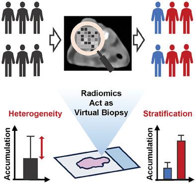
Accurately predicting nanomedicine accumulation is critical for guiding patient stratification and optimizing treatment strategies in the context of precision medicine. However, non-invasive prediction of nanomedicine accumulation remains challenging, primarily due to the complexity of identifying relevant imaging features that predict accumulation. Here, a novel non-invasive method is proposed that utilizes standard-of-care medical imaging modalities, including computed tomography and ultrasound, combined with a radiomics-based model to predict nanomedicine accumulation in tumor. The model is validated using a test dataset consisting of seven tumor xenografts in mice and three sizes of gold nanoparticles, achieving an area under the receiver operating characteristic curve of 0.851. The median accumulation levels of tumors predicted as “high accumulators” are 2.69 times greater than those predicted as “low accumulators”. Analysis of this machine-learning-driven interpretable radiomics model revealed imaging features that are strongly correlated with dense stroma, a recognized biological barrier to effective nanomedicine delivery. Radiomics-based prediction of tumor accumulation holds promise for stratifying patient and enabling precise tailoring of nanomedicine treatment strategies.
期刊介绍:
Advanced Materials, one of the world's most prestigious journals and the foundation of the Advanced portfolio, is the home of choice for best-in-class materials science for more than 30 years. Following this fast-growing and interdisciplinary field, we are considering and publishing the most important discoveries on any and all materials from materials scientists, chemists, physicists, engineers as well as health and life scientists and bringing you the latest results and trends in modern materials-related research every week.

 求助内容:
求助内容: 应助结果提醒方式:


