Claudia Carlantoni, Leon M H Liekfeld, Manu Beerens, Maike Frye
{"title":"Same same but different? How blood and lymphatic vessels induce cell contact inhibition.","authors":"Claudia Carlantoni, Leon M H Liekfeld, Manu Beerens, Maike Frye","doi":"10.1042/BST20240573","DOIUrl":null,"url":null,"abstract":"<p><p>Endothelial cells (ECs) migrate, sprout, and proliferate in response to (lymph)angiogenic mitogens, such as vascular endothelial growth factors. When ECs reach high confluency and encounter spatial confinement, they establish mature cell-cell junctions, reduce proliferation, and enter a quiescent state through a process known as contact inhibition. However, EC quiescence is modulated not only by spatial confinement but also by other mechano-environmental factors, including blood or lymph flow and extracellular matrix properties. Changes in physical forces and intracellular signaling can disrupt contact inhibition, resulting in aberrant proliferation and vascular dysfunction. Therefore, it is critical to understand the mechanisms by which endothelial cells regulate contact inhibition. While contact inhibition has been well studied in blood endothelial cells (BECs), its regulation in lymphatic endothelial cells (LECs) remains largely unexplored. Here, we review the current knowledge on extrinsic stimuli and intrinsic molecular pathways that govern endothelial contact inhibition and highlight nuanced differences between BECs and LECs. Furthermore, we provide perspectives for future research on lymphatic contact inhibition. A deeper understanding of the BEC and LEC-specific pathways underlying contact inhibition may enable targeted modulation of this process in blood or lymphatic vessels with relevance to lymphatic or blood vascular-specific disorders.</p>","PeriodicalId":8841,"journal":{"name":"Biochemical Society transactions","volume":"53 1","pages":""},"PeriodicalIF":4.3000,"publicationDate":"2025-02-06","publicationTypes":"Journal Article","fieldsOfStudy":null,"isOpenAccess":false,"openAccessPdf":"https://www.ncbi.nlm.nih.gov/pmc/articles/PMC12224917/pdf/","citationCount":"0","resultStr":null,"platform":"Semanticscholar","paperid":null,"PeriodicalName":"Biochemical Society transactions","FirstCategoryId":"99","ListUrlMain":"https://doi.org/10.1042/BST20240573","RegionNum":3,"RegionCategory":"生物学","ArticlePicture":[],"TitleCN":null,"AbstractTextCN":null,"PMCID":null,"EPubDate":"","PubModel":"","JCR":"Q2","JCRName":"BIOCHEMISTRY & MOLECULAR BIOLOGY","Score":null,"Total":0}
引用次数: 0
Abstract
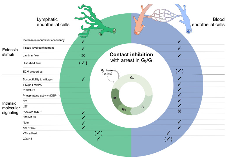
Endothelial cells (ECs) migrate, sprout, and proliferate in response to (lymph)angiogenic mitogens, such as vascular endothelial growth factors. When ECs reach high confluency and encounter spatial confinement, they establish mature cell-cell junctions, reduce proliferation, and enter a quiescent state through a process known as contact inhibition. However, EC quiescence is modulated not only by spatial confinement but also by other mechano-environmental factors, including blood or lymph flow and extracellular matrix properties. Changes in physical forces and intracellular signaling can disrupt contact inhibition, resulting in aberrant proliferation and vascular dysfunction. Therefore, it is critical to understand the mechanisms by which endothelial cells regulate contact inhibition. While contact inhibition has been well studied in blood endothelial cells (BECs), its regulation in lymphatic endothelial cells (LECs) remains largely unexplored. Here, we review the current knowledge on extrinsic stimuli and intrinsic molecular pathways that govern endothelial contact inhibition and highlight nuanced differences between BECs and LECs. Furthermore, we provide perspectives for future research on lymphatic contact inhibition. A deeper understanding of the BEC and LEC-specific pathways underlying contact inhibition may enable targeted modulation of this process in blood or lymphatic vessels with relevance to lymphatic or blood vascular-specific disorders.
期刊介绍:
Biochemical Society Transactions is the reviews journal of the Biochemical Society. Publishing concise reviews written by experts in the field, providing a timely snapshot of the latest developments across all areas of the molecular and cellular biosciences.
Elevating our authors’ ideas and expertise, each review includes a perspectives section where authors offer comment on the latest advances, a glimpse of future challenges and highlighting the importance of associated research areas in far broader contexts.
 求助内容:
求助内容: 应助结果提醒方式:


