Tubular ER structures shaped by ER-phagy receptors engage in stress-induced Golgi bypass
IF 10.7
1区 生物学
Q1 CELL BIOLOGY
引用次数: 0
Abstract
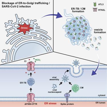
Cellular stresses, particularly endoplasmic reticulum (ER) stress induced by ER-to-Golgi transport blockade, trigger Golgi-independent secretion of cytosolic and transmembrane proteins. However, the molecular mechanisms underlying this unconventional protein secretion (UPS) remain largely elusive. Here, we report that an ER tubulovesicular structure (ER tubular body [ER-TB]), shaped by the tubular ER-phagy receptors ATL3 and RTN3L, plays an important role in stress-induced UPS of transmembrane proteins such as cystic fibrosis transmembrane conductance regulator (CFTR) and severe acute respiratory syndrome coronavirus 2 (SARS-CoV-2) spike protein. Correlative light-electron microscopy analyses demonstrate the formation of ER-TB under UPS-inducing conditions in HEK293 and HeLa cells. Individual gene knockdowns of ATL3 and RTN3 inhibit ER-TB formation and the UPS of trafficking-deficient ΔF508-CFTR. Combined supplementation of ATL3 and RTN3L induces ER-TB formation and UPS. ATL3 also participates in the SARS-CoV-2-associated convoluted membrane formation and Golgi-independent trafficking of SARS-CoV-2 spike protein. These findings suggest that ER-TB serves a common function in mediating stress-induced UPS, which participates in various physiological and pathophysiological processes.
求助全文
约1分钟内获得全文
求助全文
来源期刊
Developmental cell
生物-发育生物学
CiteScore
18.90
自引率
1.70%
发文量
203
审稿时长
3-6 weeks
期刊介绍:
Developmental Cell, established in 2001, is a comprehensive journal that explores a wide range of topics in cell and developmental biology. Our publication encompasses work across various disciplines within biology, with a particular emphasis on investigating the intersections between cell biology, developmental biology, and other related fields. Our primary objective is to present research conducted through a cell biological perspective, addressing the essential mechanisms governing cell function, cellular interactions, and responses to the environment. Moreover, we focus on understanding the collective behavior of cells, culminating in the formation of tissues, organs, and whole organisms, while also investigating the consequences of any malfunctions in these intricate processes.
 求助内容:
求助内容: 应助结果提醒方式:


