Eya Slama, S Ben Hamida, S Zaouga, H Elloumi, I Cheikh, A G Dahak, M Missaoui, A Chelibi, I Ganzoui
{"title":"Primary adenocarcinoma of duodenal bulb mimicking a pancreatic tumor: a case report.","authors":"Eya Slama, S Ben Hamida, S Zaouga, H Elloumi, I Cheikh, A G Dahak, M Missaoui, A Chelibi, I Ganzoui","doi":"10.1080/20565623.2025.2456651","DOIUrl":null,"url":null,"abstract":"<p><p>Duodenal bulb adenocarcinoma is a rare malignancy of the gastrointestinal tract. This cancer often presents with an insidious onset, making it challenging to diagnose early. A noteworthy case-study involved a 56 year-old-woman complaining from epigastric transfixing pain, jaundice, and overall health decline. The initial diagnosis suspected was a pancreatic tumor. An abdominal CT scan however showed no abnormality in the pancreas but revealed on the other hand an irregular, non-stenosing, heterogeneous fibrous thickening of the lower aspect of the duodenal bulb. An EGD showed a congested infiltrated area in the post-bulbar region, and Pathological exam confirmed the diagnosis of a primary adenocarcinoma of the duodenal bulb. The purpose of this article is to present the case of a primary adenocarcinoma of duodenal bulb mimicking a pancreatic tumor: a rare presentation of a rare tumor itself and to present points differentiating these two neoplasms. Diagnosis relies on a comprehensive assessment involving clinical, radiological, endoscopic, and histologic features. Integration of advanced imaging modalities, such as CT/MRI, coupled with upper gastrointestinal endoscopy, is crucial for accurate evaluation.</p>","PeriodicalId":12568,"journal":{"name":"Future Science OA","volume":"11 1","pages":"2456651"},"PeriodicalIF":2.1000,"publicationDate":"2025-12-01","publicationTypes":"Journal Article","fieldsOfStudy":null,"isOpenAccess":false,"openAccessPdf":"https://www.ncbi.nlm.nih.gov/pmc/articles/PMC11801341/pdf/","citationCount":"0","resultStr":null,"platform":"Semanticscholar","paperid":null,"PeriodicalName":"Future Science OA","FirstCategoryId":"1085","ListUrlMain":"https://doi.org/10.1080/20565623.2025.2456651","RegionNum":0,"RegionCategory":null,"ArticlePicture":[],"TitleCN":null,"AbstractTextCN":null,"PMCID":null,"EPubDate":"2025/2/4 0:00:00","PubModel":"Epub","JCR":"Q3","JCRName":"MEDICINE, RESEARCH & EXPERIMENTAL","Score":null,"Total":0}
引用次数: 0
Abstract
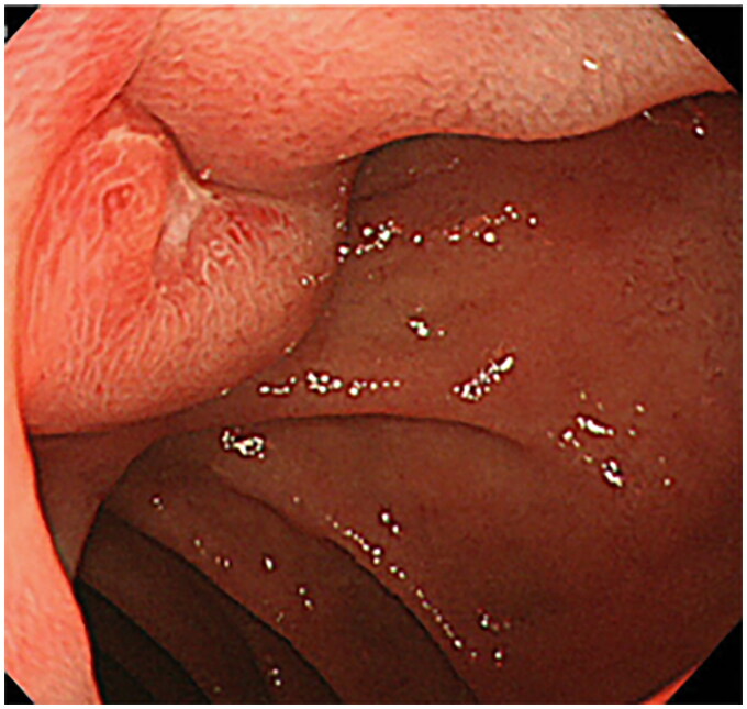
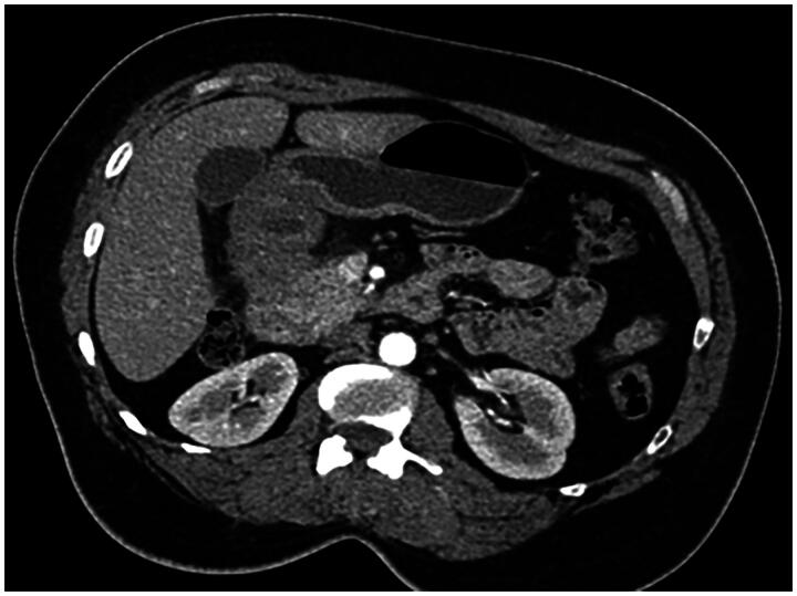
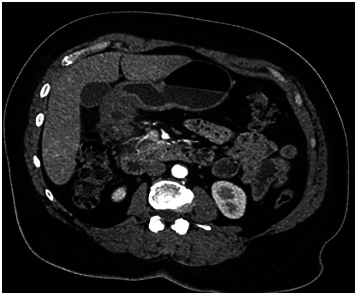
Duodenal bulb adenocarcinoma is a rare malignancy of the gastrointestinal tract. This cancer often presents with an insidious onset, making it challenging to diagnose early. A noteworthy case-study involved a 56 year-old-woman complaining from epigastric transfixing pain, jaundice, and overall health decline. The initial diagnosis suspected was a pancreatic tumor. An abdominal CT scan however showed no abnormality in the pancreas but revealed on the other hand an irregular, non-stenosing, heterogeneous fibrous thickening of the lower aspect of the duodenal bulb. An EGD showed a congested infiltrated area in the post-bulbar region, and Pathological exam confirmed the diagnosis of a primary adenocarcinoma of the duodenal bulb. The purpose of this article is to present the case of a primary adenocarcinoma of duodenal bulb mimicking a pancreatic tumor: a rare presentation of a rare tumor itself and to present points differentiating these two neoplasms. Diagnosis relies on a comprehensive assessment involving clinical, radiological, endoscopic, and histologic features. Integration of advanced imaging modalities, such as CT/MRI, coupled with upper gastrointestinal endoscopy, is crucial for accurate evaluation.
期刊介绍:
Future Science OA is an online, open access, peer-reviewed title from the Future Science Group. The journal covers research and discussion related to advances in biotechnology, medicine and health. The journal embraces the importance of publishing all good-quality research with the potential to further the progress of research in these fields. All original research articles will be considered that are within the journal''s scope, and have been conducted with scientific rigour and research integrity. The journal also features review articles, editorials and perspectives, providing readers with a leading source of commentary and analysis. Submissions of the following article types will be considered: -Research articles -Preliminary communications -Short communications -Methodologies -Trial design articles -Trial results (including early-phase and negative studies) -Reviews -Perspectives -Commentaries

 求助内容:
求助内容: 应助结果提醒方式:


