Diagnosis of Colorectal Cancer and Adenomatous Polyps of the Colon Based on the Level of MicroRNA Expression in the Mucous Membrane (Pilot Clinical Study).
M V Bagryantsev, A A Yanyshev, M G Ryabkov, A I Abelevich, I L Dezortsev, A V Bazayev
{"title":"Diagnosis of Colorectal Cancer and Adenomatous Polyps of the Colon Based on the Level of MicroRNA Expression in the Mucous Membrane (Pilot Clinical Study).","authors":"M V Bagryantsev, A A Yanyshev, M G Ryabkov, A I Abelevich, I L Dezortsev, A V Bazayev","doi":"10.17691/stm2024.16.5.05","DOIUrl":null,"url":null,"abstract":"<p><p>The gold standard for colorectal cancer (CRC) diagnosis, colonoscopy with biopsy, is an invasive technique and has some limitations, while the known non-invasive methods do not possess sufficient sensitivity and specificity. The application of microRNA as a diagnostic and prognostic CRC biomarker may compensate for the colonoscopy limitations. However, there are no data in the literature on the existence of real test systems based on the evaluation of microRNA expression. Our pilot study is the first step to creating a test system for CRC diagnosis based on the analysis of microRNA expression in the tissue of the intact colon. <b>The aim of the study</b> is to assess the prospects of using the level of microRNA expression as a supplemental method for diagnosing colorectal cancer and adenomatous polyps.</p><p><strong>Materials and methods: </strong>Patients participating in the study were divided into three groups: group 1 included patients with CRC (n=5), group 2 - patients with polyps in the colon (n=4), and patients without oncological pathology treated for hemorrhoidal disease without exacerbation (n=5) composed group 3.Tissue samples of the intact gut were taken from all patients. In groups 1 and 2, biopsy was performed in the process of right-sided laparoscopic resection of the colon with tumor. Samples of the mucous membrane from the distal part of the rectum in patients of group 3 were also collected intraoperatively; they were operated on using Milligan-Morgan technique. In groups 1 and 2, CRC and polyp samples, respectively, were taken for the analysis additionally to the intact gut area.The test panel included the following microRNAs: hsa-miR-10b-5p, hsa-miR-20a-5p, hsa-miR-141-3p, hsa-miR-181b-5p. The levels of the reference genes were analyzed with the help of real-time polymerase chain reaction using intercalating SYBR Green stain.</p><p><strong>Results: </strong>Expression of hsa-miR-141-3p in the mucous membrane of the colon in patients of groups 1 and 2 (with CRC and polyps, respectively) was statistically significantly higher than in patients without bowel tumors. At the same time, the expression level of hsa-miR-10b-5p was statistically significantly lower in the tumor tissue (cancer or polyps) in comparison with patients of group 3.Lower values of expression in all tested microRNAs have been detected in the CRC tissue relative to the intact mucosa of the same patients. A similar tendency was also observed in patients with adenomatous polyps.</p><p><strong>Conclusion: </strong>The results of the study have shown that of four microRNAs, included into the test panel, hsa-miR-141-3p and hsa-miR-10b-5p were found to have the diagnostic value for identifying tumor colorectal lesions. Thus, our data will assume that supplementing the endoscopic tests of the large intestine by the epigenetic analysis of the mucous membrane is a promising approach to cancer screening procedures.</p>","PeriodicalId":520289,"journal":{"name":"Sovremennye tekhnologii v meditsine","volume":"16 5","pages":"45-51"},"PeriodicalIF":0.0000,"publicationDate":"2024-01-01","publicationTypes":"Journal Article","fieldsOfStudy":null,"isOpenAccess":false,"openAccessPdf":"https://www.ncbi.nlm.nih.gov/pmc/articles/PMC11784883/pdf/","citationCount":"0","resultStr":null,"platform":"Semanticscholar","paperid":null,"PeriodicalName":"Sovremennye tekhnologii v meditsine","FirstCategoryId":"1085","ListUrlMain":"https://doi.org/10.17691/stm2024.16.5.05","RegionNum":0,"RegionCategory":null,"ArticlePicture":[],"TitleCN":null,"AbstractTextCN":null,"PMCID":null,"EPubDate":"2024/10/30 0:00:00","PubModel":"Epub","JCR":"","JCRName":"","Score":null,"Total":0}
引用次数: 0
Abstract
The gold standard for colorectal cancer (CRC) diagnosis, colonoscopy with biopsy, is an invasive technique and has some limitations, while the known non-invasive methods do not possess sufficient sensitivity and specificity. The application of microRNA as a diagnostic and prognostic CRC biomarker may compensate for the colonoscopy limitations. However, there are no data in the literature on the existence of real test systems based on the evaluation of microRNA expression. Our pilot study is the first step to creating a test system for CRC diagnosis based on the analysis of microRNA expression in the tissue of the intact colon. The aim of the study is to assess the prospects of using the level of microRNA expression as a supplemental method for diagnosing colorectal cancer and adenomatous polyps.
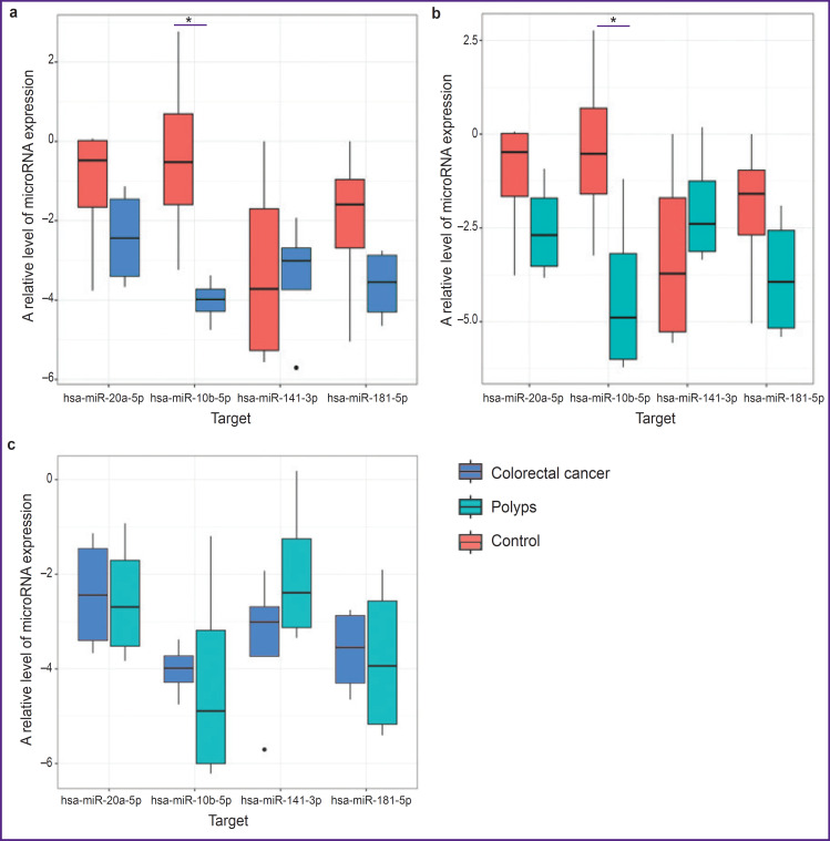
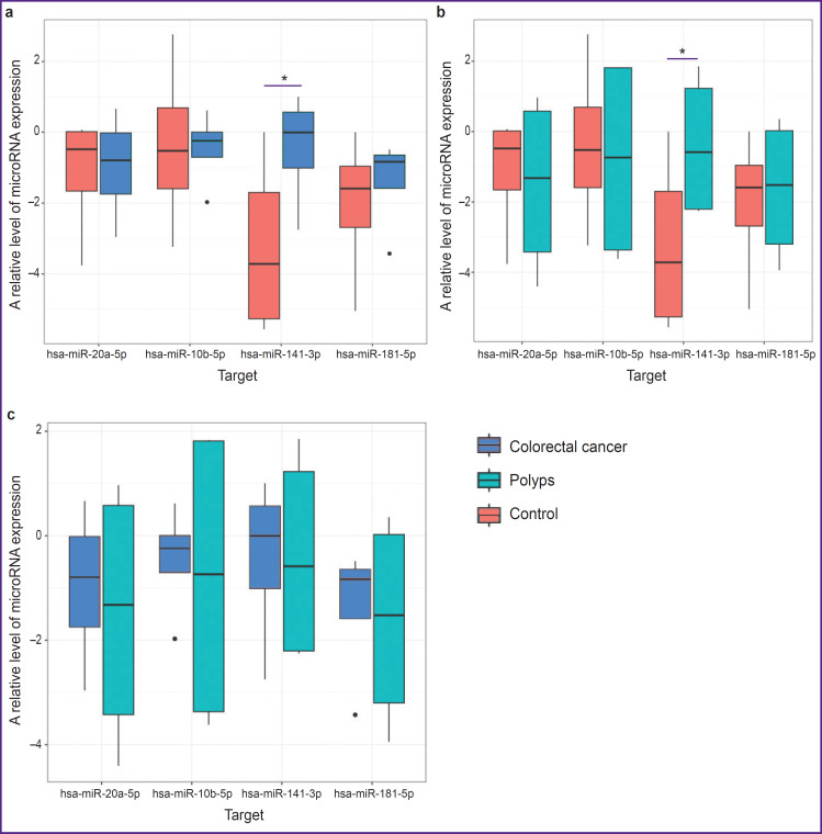
Materials and methods: Patients participating in the study were divided into three groups: group 1 included patients with CRC (n=5), group 2 - patients with polyps in the colon (n=4), and patients without oncological pathology treated for hemorrhoidal disease without exacerbation (n=5) composed group 3.Tissue samples of the intact gut were taken from all patients. In groups 1 and 2, biopsy was performed in the process of right-sided laparoscopic resection of the colon with tumor. Samples of the mucous membrane from the distal part of the rectum in patients of group 3 were also collected intraoperatively; they were operated on using Milligan-Morgan technique. In groups 1 and 2, CRC and polyp samples, respectively, were taken for the analysis additionally to the intact gut area.The test panel included the following microRNAs: hsa-miR-10b-5p, hsa-miR-20a-5p, hsa-miR-141-3p, hsa-miR-181b-5p. The levels of the reference genes were analyzed with the help of real-time polymerase chain reaction using intercalating SYBR Green stain.
Results: Expression of hsa-miR-141-3p in the mucous membrane of the colon in patients of groups 1 and 2 (with CRC and polyps, respectively) was statistically significantly higher than in patients without bowel tumors. At the same time, the expression level of hsa-miR-10b-5p was statistically significantly lower in the tumor tissue (cancer or polyps) in comparison with patients of group 3.Lower values of expression in all tested microRNAs have been detected in the CRC tissue relative to the intact mucosa of the same patients. A similar tendency was also observed in patients with adenomatous polyps.
Conclusion: The results of the study have shown that of four microRNAs, included into the test panel, hsa-miR-141-3p and hsa-miR-10b-5p were found to have the diagnostic value for identifying tumor colorectal lesions. Thus, our data will assume that supplementing the endoscopic tests of the large intestine by the epigenetic analysis of the mucous membrane is a promising approach to cancer screening procedures.

 求助内容:
求助内容: 应助结果提醒方式:


