Echocardiographic Evaluation of Central Venous Pressure Using Inferior Vena Cava Characteristics: An Estimate Guide for Right Atrial Pressure in Intensive Care Unit.
Muataz F Hussein, Wisam J Mohammad, Samar Omran Essa
{"title":"Echocardiographic Evaluation of Central Venous Pressure Using Inferior Vena Cava Characteristics: An Estimate Guide for Right Atrial Pressure in Intensive Care Unit.","authors":"Muataz F Hussein, Wisam J Mohammad, Samar Omran Essa","doi":"10.4103/jcecho.jcecho_2_24","DOIUrl":null,"url":null,"abstract":"<p><strong>Background: </strong>Central venous pressure (CVP) is a good approximation of right atrial pressure (RAP), which in turn is a major determinant of right ventricular filling. The inferior vena cava (IVC) is a compliant vessel whose size and shape vary with changes in CVP. IVC diameter and Collapsibility Index (CI) assessed by echocardiography are used as indirect indicators for the estimation of RAP.</p><p><strong>Aim of the study: </strong>To evaluate the correlation between IVC echocardiographic characteristics and CVP and RAP and the value of assessment of IVC as a guide for the status of the right side of the heart.</p><p><strong>Patients and methods: </strong>A total of sixty patients (male and female) above 18 years of age, who were admitted in the intensive care unit, were enrolled in this single-center, descriptive cross-sectional study. Echocardiographic assessment of IVC hemodynamics (IVC expiratory [IVCe] and inspiratory [IVCi] diameters and IVC-CI) were carried out. In addition to standard echocardiographic examination, right heart function measurements (Tricuspid annular plane systolic excursion [TAPSE] and right atrial [RA] area) in spontaneously and mechanically ventilated patients were done.</p><p><strong>Results: </strong>The average age of the patients was 62 years (18-80 years). Overall, 45% (<i>n</i> = 27) were male and 55% (<i>n</i> = 33) were female. The breathing modality was mechanical ventilation in 27 (45%) patients and spontaneous breathing in 33 (55%) patients. Both IVCe and IVCi diameters showed a strong negative correlation with CI, (<i>r</i> = -0.920 for IVCe and <i>r</i> = -0.964 for IVCi) (<i>P</i> < 0.001). There was a positive correlation between TAPSE and IVC-CI (<i>r</i> = 0.857, <i>P</i> < 0.001). IVC-CI in mechanically ventilated patients was (mean ± standard deviation [SD], 40.11 ± 1.782) compared to spontaneous breathing (mean ± SD, 48.91 ± 1.811) (<i>P</i> < 0.001).</p><p><strong>Conclusions: </strong>There is a linear relationship of IVC-CI with TAPSE but an inverse relation with RA area. Evaluation of IVC diameter and its CI is an easy and noninvasive method to estimate CVP and RAP and so evaluate right heart performance of critically ill patients. Its use is more helpful in patients who are spontaneously breathing than those who are mechanically ventilated.</p>","PeriodicalId":15191,"journal":{"name":"Journal of Cardiovascular Echography","volume":"34 4","pages":"206-213"},"PeriodicalIF":1.0000,"publicationDate":"2024-10-01","publicationTypes":"Journal Article","fieldsOfStudy":null,"isOpenAccess":false,"openAccessPdf":"https://www.ncbi.nlm.nih.gov/pmc/articles/PMC11784733/pdf/","citationCount":"0","resultStr":null,"platform":"Semanticscholar","paperid":null,"PeriodicalName":"Journal of Cardiovascular Echography","FirstCategoryId":"1085","ListUrlMain":"https://doi.org/10.4103/jcecho.jcecho_2_24","RegionNum":0,"RegionCategory":null,"ArticlePicture":[],"TitleCN":null,"AbstractTextCN":null,"PMCID":null,"EPubDate":"2024/12/19 0:00:00","PubModel":"Epub","JCR":"Q4","JCRName":"CARDIAC & CARDIOVASCULAR SYSTEMS","Score":null,"Total":0}
引用次数: 0
Abstract
Background: Central venous pressure (CVP) is a good approximation of right atrial pressure (RAP), which in turn is a major determinant of right ventricular filling. The inferior vena cava (IVC) is a compliant vessel whose size and shape vary with changes in CVP. IVC diameter and Collapsibility Index (CI) assessed by echocardiography are used as indirect indicators for the estimation of RAP.
Aim of the study: To evaluate the correlation between IVC echocardiographic characteristics and CVP and RAP and the value of assessment of IVC as a guide for the status of the right side of the heart.
Patients and methods: A total of sixty patients (male and female) above 18 years of age, who were admitted in the intensive care unit, were enrolled in this single-center, descriptive cross-sectional study. Echocardiographic assessment of IVC hemodynamics (IVC expiratory [IVCe] and inspiratory [IVCi] diameters and IVC-CI) were carried out. In addition to standard echocardiographic examination, right heart function measurements (Tricuspid annular plane systolic excursion [TAPSE] and right atrial [RA] area) in spontaneously and mechanically ventilated patients were done.
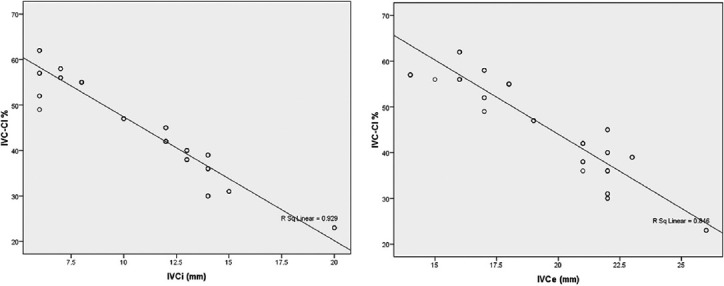
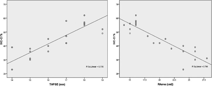
Results: The average age of the patients was 62 years (18-80 years). Overall, 45% (n = 27) were male and 55% (n = 33) were female. The breathing modality was mechanical ventilation in 27 (45%) patients and spontaneous breathing in 33 (55%) patients. Both IVCe and IVCi diameters showed a strong negative correlation with CI, (r = -0.920 for IVCe and r = -0.964 for IVCi) (P < 0.001). There was a positive correlation between TAPSE and IVC-CI (r = 0.857, P < 0.001). IVC-CI in mechanically ventilated patients was (mean ± standard deviation [SD], 40.11 ± 1.782) compared to spontaneous breathing (mean ± SD, 48.91 ± 1.811) (P < 0.001).
Conclusions: There is a linear relationship of IVC-CI with TAPSE but an inverse relation with RA area. Evaluation of IVC diameter and its CI is an easy and noninvasive method to estimate CVP and RAP and so evaluate right heart performance of critically ill patients. Its use is more helpful in patients who are spontaneously breathing than those who are mechanically ventilated.

 求助内容:
求助内容: 应助结果提醒方式:


