Lauren B Yeager, Chloe Y Li, Dmitry Bogolmony, Larisa Debelenko, Brian P Marr
{"title":"Vitreous Hemorrhage in Posterior Uveal Melanocytoma: Two Case Reports.","authors":"Lauren B Yeager, Chloe Y Li, Dmitry Bogolmony, Larisa Debelenko, Brian P Marr","doi":"10.1155/crop/5126550","DOIUrl":null,"url":null,"abstract":"<p><p><b>Purpose:</b> We describe the diagnostic challenge by melanocytomas originating from locations other than the optic nerve. <b>Methods:</b> This is a retrospective case report of two patients who presented with vitreous hemorrhage with underlying melanocytomas. <b>Results:</b> Both patients had hemorrhage obscuring indeterminant uveal masses; fine-needle aspiration biopsies confirmed melanocytoma with necrosis and atypia and, in one case, concern for malignant transformation. <b>Conclusion:</b> Melanocytomas are rare, benign melanocytic lesions that resemble nevi. In contrast to optic nerve melanocytoma, those involving the choroid and ciliary body lack specific clinical characteristics. Vitreous hemorrhage is an underrecognized complication, and uveal melanocytoma must be included in the differential diagnosis of vitreous hemorrhage with associated ciliary body or choroidal mass. Biopsy is required for definitive diagnosis and to identify malignant transformation of the lesion, a rare but possible occurrence.</p>","PeriodicalId":9603,"journal":{"name":"Case Reports in Ophthalmological Medicine","volume":"2025 ","pages":"5126550"},"PeriodicalIF":0.4000,"publicationDate":"2025-01-22","publicationTypes":"Journal Article","fieldsOfStudy":null,"isOpenAccess":false,"openAccessPdf":"https://www.ncbi.nlm.nih.gov/pmc/articles/PMC11779989/pdf/","citationCount":"0","resultStr":null,"platform":"Semanticscholar","paperid":null,"PeriodicalName":"Case Reports in Ophthalmological Medicine","FirstCategoryId":"1085","ListUrlMain":"https://doi.org/10.1155/crop/5126550","RegionNum":0,"RegionCategory":null,"ArticlePicture":[],"TitleCN":null,"AbstractTextCN":null,"PMCID":null,"EPubDate":"2025/1/1 0:00:00","PubModel":"eCollection","JCR":"Q4","JCRName":"OPHTHALMOLOGY","Score":null,"Total":0}
引用次数: 0
Abstract
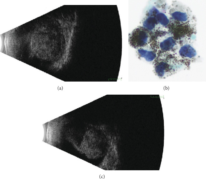
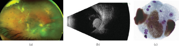
Purpose: We describe the diagnostic challenge by melanocytomas originating from locations other than the optic nerve. Methods: This is a retrospective case report of two patients who presented with vitreous hemorrhage with underlying melanocytomas. Results: Both patients had hemorrhage obscuring indeterminant uveal masses; fine-needle aspiration biopsies confirmed melanocytoma with necrosis and atypia and, in one case, concern for malignant transformation. Conclusion: Melanocytomas are rare, benign melanocytic lesions that resemble nevi. In contrast to optic nerve melanocytoma, those involving the choroid and ciliary body lack specific clinical characteristics. Vitreous hemorrhage is an underrecognized complication, and uveal melanocytoma must be included in the differential diagnosis of vitreous hemorrhage with associated ciliary body or choroidal mass. Biopsy is required for definitive diagnosis and to identify malignant transformation of the lesion, a rare but possible occurrence.

 求助内容:
求助内容: 应助结果提醒方式:


