Ischemic stroke reduces bone perfusion and alters osteovascular structure
IF 2.6
Q3 ENDOCRINOLOGY & METABOLISM
引用次数: 0
Abstract
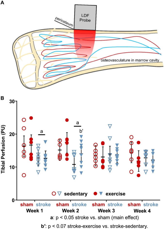
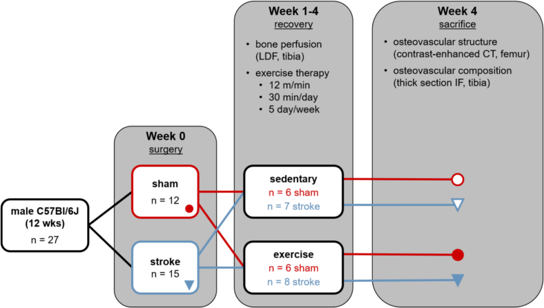
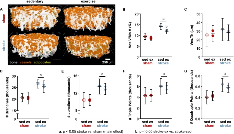
Stroke patients lose bone mass and experience fracture at an elevated rate. Although functional intraosseous vasculature is necessary for skeletal maintenance, the effect of stroke on osteovasculature is unknown. In this study we characterized changes to osteovascular perfusion, structure, and composition following mild-to-moderate stroke severity in mice, both with and without exercise therapy. Twelve-week-old male mice (n = 27) received either an ischemic stroke (middle cerebral artery occlusion) or sham procedure, followed by a four-week recovery with either moderate daily treadmill or sedentary activity. Intraosseous perfusion, measured weekly in the proximal tibial metaphysis with laser Doppler flowmetry, was reduced for two weeks in the stroke group relative to the sham group. After four weeks, osteovascular structure was assessed in the distal femoral metaphysis with contrast-enhanced computed tomography. Increased osteovascular volume and branching, decreased number of smaller vessels (6–22 μm), and increased number of larger vessels (>66 μm) were observed in the stroke groups compared to sham groups, which may be a compensatory response to early perfusion deficits. Although moderate exercise mitigated the impact of stroke on osteovascular perfusion and volume, it tended to reduce the amount of osteogenic type H vasculature quantified with immunofluorescence microscopy and, exacerbated by stroke effects, produced fewer vessels in close proximity to bone and thus may have detrimental effects on bone remodeling during early stroke recovery. Since results were similar in both limbs, the effects of ischemic stroke on osteovascular perfusion and structure were primarily systemic, rather than resulting from paresis or disuse, providing new insight for future studies on the pathogenesis and treatment of skeletal fragility in stroke patients.
缺血性中风减少骨灌注,改变骨血管结构。
中风患者骨量减少,骨折发生率升高。虽然骨内血管系统的功能是骨骼维持所必需的,但中风对骨内血管系统的影响尚不清楚。在这项研究中,我们描述了在小鼠轻度至中度中风严重程度后,无论是否进行运动治疗,骨血管灌注、结构和组成的变化。12周大的雄性小鼠(n = 27)接受缺血性中风(大脑中动脉闭塞)或假手术,随后进行为期四周的恢复,每天进行适度的跑步机或久坐活动。用激光多普勒血流仪每周测量胫骨近端干骺端骨内灌注,与假手术组相比,中风组的骨内灌注减少了两周。四周后,通过增强计算机断层扫描评估股骨远端干骺端骨血管结构。与假手术组相比,中风组骨血管体积和分支增加,小血管(6-22 μm)数量减少,大血管(> - 66 μm)数量增加,这可能是对早期灌注缺陷的代偿反应。虽然适度运动减轻了中风对骨血管灌注和体积的影响,但它倾向于减少免疫荧光显微镜量化的成骨H型血管的数量,并且由于中风的影响而加剧,在靠近骨骼的地方产生更少的血管,因此可能对早期中风恢复期间的骨重塑产生不利影响。由于两肢结果相似,缺血性脑卒中对骨血管灌注和结构的影响主要是全身性的,而不是由轻瘫或废用引起的,这为未来脑卒中患者骨骼脆性的发病机制和治疗研究提供了新的思路。
本文章由计算机程序翻译,如有差异,请以英文原文为准。
求助全文
约1分钟内获得全文
求助全文
来源期刊
Bone Reports
Medicine-Orthopedics and Sports Medicine
CiteScore
4.30
自引率
4.00%
发文量
444
审稿时长
57 days
期刊介绍:
Bone Reports is an interdisciplinary forum for the rapid publication of Original Research Articles and Case Reports across basic, translational and clinical aspects of bone and mineral metabolism. The journal publishes papers that are scientifically sound, with the peer review process focused principally on verifying sound methodologies, and correct data analysis and interpretation. We welcome studies either replicating or failing to replicate a previous study, and null findings. We fulfil a critical and current need to enhance research by publishing reproducibility studies and null findings.
 求助内容:
求助内容: 应助结果提醒方式:


