Karl D. Briegel, Nick R. von Grafenstein, Julia C. Draeger, Peter Blümler, Robin D. Allert, Dominik B. Bucher
{"title":"Optical widefield nuclear magnetic resonance microscopy","authors":"Karl D. Briegel, Nick R. von Grafenstein, Julia C. Draeger, Peter Blümler, Robin D. Allert, Dominik B. Bucher","doi":"10.1038/s41467-024-55003-5","DOIUrl":null,"url":null,"abstract":"<p>Microscopy enables detailed visualization and understanding of minute structures or processes. While cameras have significantly advanced optical, infrared, and electron microscopy, imaging nuclear magnetic resonance (NMR) signals on a camera has remained elusive. Here, we employ nitrogen-vacancy centers in diamond as a quantum sensor, which converts NMR signals into optical signals that are subsequently captured by a high-speed camera. Unlike traditional magnetic resonance imaging, our method records the NMR signal over a wide field of view in real space. We demonstrate that our optical widefield NMR microscopy can image NMR signals in microfluidic structures with a ~10 μm resolution across a ~235 × 150 μm<sup>2</sup> area. Crucially, each camera pixel records an NMR spectrum providing multicomponent information about the signal’s amplitude, phase, local magnetic field strengths, and gradients. The fusion of optical microscopy and NMR techniques enables multifaceted imaging applications in the physical and life sciences.</p>","PeriodicalId":19066,"journal":{"name":"Nature Communications","volume":"122 1","pages":""},"PeriodicalIF":15.7000,"publicationDate":"2025-02-03","publicationTypes":"Journal Article","fieldsOfStudy":null,"isOpenAccess":false,"openAccessPdf":"","citationCount":"0","resultStr":null,"platform":"Semanticscholar","paperid":null,"PeriodicalName":"Nature Communications","FirstCategoryId":"103","ListUrlMain":"https://doi.org/10.1038/s41467-024-55003-5","RegionNum":1,"RegionCategory":"综合性期刊","ArticlePicture":[],"TitleCN":null,"AbstractTextCN":null,"PMCID":null,"EPubDate":"","PubModel":"","JCR":"Q1","JCRName":"MULTIDISCIPLINARY SCIENCES","Score":null,"Total":0}
引用次数: 0
Abstract
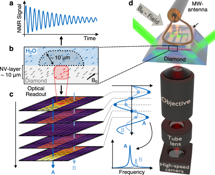
Microscopy enables detailed visualization and understanding of minute structures or processes. While cameras have significantly advanced optical, infrared, and electron microscopy, imaging nuclear magnetic resonance (NMR) signals on a camera has remained elusive. Here, we employ nitrogen-vacancy centers in diamond as a quantum sensor, which converts NMR signals into optical signals that are subsequently captured by a high-speed camera. Unlike traditional magnetic resonance imaging, our method records the NMR signal over a wide field of view in real space. We demonstrate that our optical widefield NMR microscopy can image NMR signals in microfluidic structures with a ~10 μm resolution across a ~235 × 150 μm2 area. Crucially, each camera pixel records an NMR spectrum providing multicomponent information about the signal’s amplitude, phase, local magnetic field strengths, and gradients. The fusion of optical microscopy and NMR techniques enables multifaceted imaging applications in the physical and life sciences.
期刊介绍:
Nature Communications, an open-access journal, publishes high-quality research spanning all areas of the natural sciences. Papers featured in the journal showcase significant advances relevant to specialists in each respective field. With a 2-year impact factor of 16.6 (2022) and a median time of 8 days from submission to the first editorial decision, Nature Communications is committed to rapid dissemination of research findings. As a multidisciplinary journal, it welcomes contributions from biological, health, physical, chemical, Earth, social, mathematical, applied, and engineering sciences, aiming to highlight important breakthroughs within each domain.

 求助内容:
求助内容: 应助结果提醒方式:


