Enhanced precision in stone localization and intraoral removal in sialolithiasis: the role of preoperative computer tomographic scanning in surgical planning.
Soo Yeon Jung, Mi Sun Chun, Yu Jin Go, Ju Hyun Yun, Han Su Kim
{"title":"Enhanced precision in stone localization and intraoral removal in sialolithiasis: the role of preoperative computer tomographic scanning in surgical planning.","authors":"Soo Yeon Jung, Mi Sun Chun, Yu Jin Go, Ju Hyun Yun, Han Su Kim","doi":"10.1186/s13005-024-00479-1","DOIUrl":null,"url":null,"abstract":"<p><strong>Background: </strong>The precise localization of stones within the submandibular duct is crucial for the successful intraoral removal in sialolithiasis. Customizing surgical approaches based on the stone's ductal location is imperative. Particularly challenging are stones beneath the lingual nerve, requiring a landmark-guided approach due to their non-palpable nature. This study aimed to comprehend stone positioning, location-specific characteristics, and develop suitable surgical approaches. We conducted a thorough analysis of numerous preoperative computed tomography (CT) scans for this purpose.</p><p><strong>Methods: </strong>We performed a retrospective review of the medical records of patients who underwent intraoral stone removal between 2006 and 2022. Two different surgical approaches were applied based on the stone location as determined by preoperative CT scans. The mediolingual approach was used for superficial stones, while the laterogingival approach was reserved for deeper stones. Patient demographics, sialolithiasis features, and postoperative complications were analyzed. T-test was performed to compare stone characteristics between different locations, and a receiver operating characteristic curve analysis was used to identify the critical size threshold for predicting stone location.</p><p><strong>Results: </strong>Medical records of 465 patients were reviewed. Out of 616 stones, 614 were successfully removed with two distinct surgical approaches guided by preoperative CT scans. Two patients reported retention, and 11 experienced postoperative tongue sensation changes. The hilum was the most common stone location, and deeper stones, approached laterolingually, were generally larger. Analysis identified a 4.25 mm width as the most sensitive and specific threshold for deep stones. Stone volume showed no statistically significant difference between smokers and non-smokers, alcohol consumers and non-consumer.</p><p><strong>Conclusion: </strong>The result of the study underscore the significance of precise stone localization and endorse the efficacy of landmark-guided surgical approaches in managing sialolithiasis.</p>","PeriodicalId":12994,"journal":{"name":"Head & Face Medicine","volume":"21 1","pages":"3"},"PeriodicalIF":2.4000,"publicationDate":"2025-01-31","publicationTypes":"Journal Article","fieldsOfStudy":null,"isOpenAccess":false,"openAccessPdf":"https://www.ncbi.nlm.nih.gov/pmc/articles/PMC11786450/pdf/","citationCount":"0","resultStr":null,"platform":"Semanticscholar","paperid":null,"PeriodicalName":"Head & Face Medicine","FirstCategoryId":"3","ListUrlMain":"https://doi.org/10.1186/s13005-024-00479-1","RegionNum":2,"RegionCategory":"医学","ArticlePicture":[],"TitleCN":null,"AbstractTextCN":null,"PMCID":null,"EPubDate":"","PubModel":"","JCR":"Q2","JCRName":"DENTISTRY, ORAL SURGERY & MEDICINE","Score":null,"Total":0}
引用次数: 0
Abstract
Background: The precise localization of stones within the submandibular duct is crucial for the successful intraoral removal in sialolithiasis. Customizing surgical approaches based on the stone's ductal location is imperative. Particularly challenging are stones beneath the lingual nerve, requiring a landmark-guided approach due to their non-palpable nature. This study aimed to comprehend stone positioning, location-specific characteristics, and develop suitable surgical approaches. We conducted a thorough analysis of numerous preoperative computed tomography (CT) scans for this purpose.
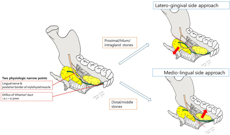
Methods: We performed a retrospective review of the medical records of patients who underwent intraoral stone removal between 2006 and 2022. Two different surgical approaches were applied based on the stone location as determined by preoperative CT scans. The mediolingual approach was used for superficial stones, while the laterogingival approach was reserved for deeper stones. Patient demographics, sialolithiasis features, and postoperative complications were analyzed. T-test was performed to compare stone characteristics between different locations, and a receiver operating characteristic curve analysis was used to identify the critical size threshold for predicting stone location.
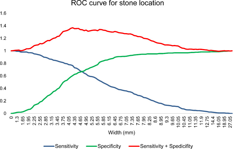
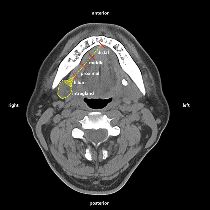
Results: Medical records of 465 patients were reviewed. Out of 616 stones, 614 were successfully removed with two distinct surgical approaches guided by preoperative CT scans. Two patients reported retention, and 11 experienced postoperative tongue sensation changes. The hilum was the most common stone location, and deeper stones, approached laterolingually, were generally larger. Analysis identified a 4.25 mm width as the most sensitive and specific threshold for deep stones. Stone volume showed no statistically significant difference between smokers and non-smokers, alcohol consumers and non-consumer.
Conclusion: The result of the study underscore the significance of precise stone localization and endorse the efficacy of landmark-guided surgical approaches in managing sialolithiasis.
期刊介绍:
Head & Face Medicine is a multidisciplinary open access journal that publishes basic and clinical research concerning all aspects of cranial, facial and oral conditions.
The journal covers all aspects of cranial, facial and oral diseases and their management. It has been designed as a multidisciplinary journal for clinicians and researchers involved in the diagnostic and therapeutic aspects of diseases which affect the human head and face. The journal is wide-ranging, covering the development, aetiology, epidemiology and therapy of head and face diseases to the basic science that underlies these diseases. Management of head and face diseases includes all aspects of surgical and non-surgical treatments including psychopharmacological therapies.
 求助内容:
求助内容: 应助结果提醒方式:


