{"title":"Virtual staining from bright-field microscopy for label-free quantitative analysis of plant cell structures.","authors":"Manami Ichita, Haruna Yamamichi, Takumi Higaki","doi":"10.1007/s11103-025-01558-w","DOIUrl":null,"url":null,"abstract":"<p><p>The applicability of a deep learning model for the virtual staining of plant cell structures using bright-field microscopy was investigated. The training dataset consisted of microscopy images of tobacco BY-2 cells with the plasma membrane stained with the fluorescent dye PlasMem Bright Green and the cell nucleus labeled with Histone-red fluorescent protein. The trained models successfully detected the expansion of cell nuclei upon aphidicolin treatment and a decrease in the cell aspect ratio upon propyzamide treatment, demonstrating its utility in cell morphometry. The model also accurately documented the shape of Arabidopsis pavement cells in both wild type and the bpp125 triple mutant, which has an altered pavement cell phenotype. Metrics such as cell area, circularity, and solidity obtained from virtual staining analyses were highly correlated with those obtained by manual measurements of cell features from microscopy images. Furthermore, the versatility of virtual staining was highlighted by its application to track chloroplast movement in Egeria densa. The method was also effective for classifying live and dead BY-2 cells using texture-based machine learning, suggesting that virtual staining can be applied beyond typical segmentation tasks. Although this method still has some limitations, its non-invasive nature and efficiency make it highly suitable for label-free, dynamic, and high-throughput analyses in quantitative plant cell biology.</p>","PeriodicalId":20064,"journal":{"name":"Plant Molecular Biology","volume":"115 1","pages":"29"},"PeriodicalIF":3.8000,"publicationDate":"2025-01-31","publicationTypes":"Journal Article","fieldsOfStudy":null,"isOpenAccess":false,"openAccessPdf":"https://www.ncbi.nlm.nih.gov/pmc/articles/PMC11782351/pdf/","citationCount":"0","resultStr":null,"platform":"Semanticscholar","paperid":null,"PeriodicalName":"Plant Molecular Biology","FirstCategoryId":"99","ListUrlMain":"https://doi.org/10.1007/s11103-025-01558-w","RegionNum":2,"RegionCategory":"生物学","ArticlePicture":[],"TitleCN":null,"AbstractTextCN":null,"PMCID":null,"EPubDate":"","PubModel":"","JCR":"Q2","JCRName":"BIOCHEMISTRY & MOLECULAR BIOLOGY","Score":null,"Total":0}
引用次数: 0
Abstract
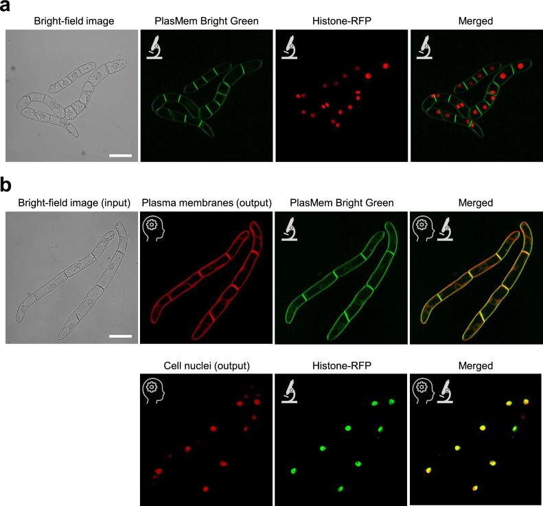
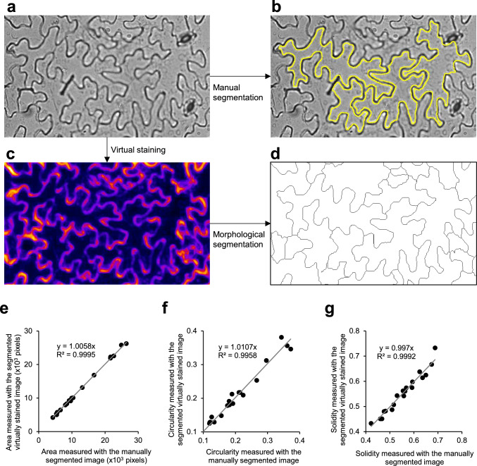
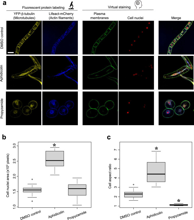
The applicability of a deep learning model for the virtual staining of plant cell structures using bright-field microscopy was investigated. The training dataset consisted of microscopy images of tobacco BY-2 cells with the plasma membrane stained with the fluorescent dye PlasMem Bright Green and the cell nucleus labeled with Histone-red fluorescent protein. The trained models successfully detected the expansion of cell nuclei upon aphidicolin treatment and a decrease in the cell aspect ratio upon propyzamide treatment, demonstrating its utility in cell morphometry. The model also accurately documented the shape of Arabidopsis pavement cells in both wild type and the bpp125 triple mutant, which has an altered pavement cell phenotype. Metrics such as cell area, circularity, and solidity obtained from virtual staining analyses were highly correlated with those obtained by manual measurements of cell features from microscopy images. Furthermore, the versatility of virtual staining was highlighted by its application to track chloroplast movement in Egeria densa. The method was also effective for classifying live and dead BY-2 cells using texture-based machine learning, suggesting that virtual staining can be applied beyond typical segmentation tasks. Although this method still has some limitations, its non-invasive nature and efficiency make it highly suitable for label-free, dynamic, and high-throughput analyses in quantitative plant cell biology.
期刊介绍:
Plant Molecular Biology is an international journal dedicated to rapid publication of original research articles in all areas of plant biology.The Editorial Board welcomes full-length manuscripts that address important biological problems of broad interest, including research in comparative genomics, functional genomics, proteomics, bioinformatics, computational biology, biochemical and regulatory networks, and biotechnology. Because space in the journal is limited, however, preference is given to publication of results that provide significant new insights into biological problems and that advance the understanding of structure, function, mechanisms, or regulation. Authors must ensure that results are of high quality and that manuscripts are written for a broad plant science audience.
 求助内容:
求助内容: 应助结果提醒方式:


