Wei Wei, Yongjun Jia, Ming Li, Nan Yu, Shan Dang, Jian Geng, Dong Han, Yong Yu, Yunsong Zheng, Lihua Fan
{"title":"Combining Low-energy Images in Dual-energy Spectral CT With Deep Learning Image Reconstruction Algorithm to Improve Inferior Vena Cava Image Quality.","authors":"Wei Wei, Yongjun Jia, Ming Li, Nan Yu, Shan Dang, Jian Geng, Dong Han, Yong Yu, Yunsong Zheng, Lihua Fan","doi":"10.1097/RCT.0000000000001713","DOIUrl":null,"url":null,"abstract":"<p><strong>Objective: </strong>To explore the application of low-energy image in dual-energy spectral CT (DEsCT) combined with deep learning image reconstruction (DLIR) to improve inferior vena cava imaging.</p><p><strong>Materials and methods: </strong>Thirty patients with inferior vena cava syndrome underwent contrast-enhanced upper abdominal CT with routine dose, and the 40, 50, 60, 70, and 80 keV images in the delayed phase were first reconstructed with the ASiR-V40% algorithm. Image quality was evaluated both quantitatively [CT value, SD, signal-to-noise ratio (SNR), and contrast-to-noise ratio (CNR) for inferior vena cava] and qualitatively to select an optimal energy level with the best image quality. Then, the optimal-energy images were reconstructed again using deep learning image reconstruction medium strength (DLIR-M) and DLIR-H (high strength) algorithms and compared with that of ASiR-V40%.</p><p><strong>Results: </strong>The objective CT value, SD, SNR, and CNR increased with the decrease in energy level, with statistically significant differences (all P <0.05). The 40 keV images had the highest CT values, SNR, and CNR and good diagnostic acceptability, and 40 keV was selected as the best energy level. Compared with ASiR-V40% and DLIR-M, DLIR-H had the lowest SD, highest SNR and CNR, and subjective score (all P <0.001) with good consistencies between the 2 physicians (all k ≥0.75). The 40 keV images with DLIR-H had the highest overall image quality, showing sharper edges of inferior vena cava vessels and clearer lumen in patients with Budd-Chiari syndrome.</p><p><strong>Conclusions: </strong>Compared with the ASiR-V algorithm, DLIR-H significantly reduces image noise and provides the highest CNR and best diagnostic image quality for the 40 keV DEsCT images in imaging inferior vena cava.</p>","PeriodicalId":15402,"journal":{"name":"Journal of Computer Assisted Tomography","volume":" ","pages":"604-610"},"PeriodicalIF":1.3000,"publicationDate":"2025-07-01","publicationTypes":"Journal Article","fieldsOfStudy":null,"isOpenAccess":false,"openAccessPdf":"https://www.ncbi.nlm.nih.gov/pmc/articles/PMC12237118/pdf/","citationCount":"0","resultStr":null,"platform":"Semanticscholar","paperid":null,"PeriodicalName":"Journal of Computer Assisted Tomography","FirstCategoryId":"3","ListUrlMain":"https://doi.org/10.1097/RCT.0000000000001713","RegionNum":4,"RegionCategory":"医学","ArticlePicture":[],"TitleCN":null,"AbstractTextCN":null,"PMCID":null,"EPubDate":"2025/1/27 0:00:00","PubModel":"Epub","JCR":"Q4","JCRName":"RADIOLOGY, NUCLEAR MEDICINE & MEDICAL IMAGING","Score":null,"Total":0}
引用次数: 0
Abstract
Objective: To explore the application of low-energy image in dual-energy spectral CT (DEsCT) combined with deep learning image reconstruction (DLIR) to improve inferior vena cava imaging.
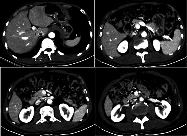
Materials and methods: Thirty patients with inferior vena cava syndrome underwent contrast-enhanced upper abdominal CT with routine dose, and the 40, 50, 60, 70, and 80 keV images in the delayed phase were first reconstructed with the ASiR-V40% algorithm. Image quality was evaluated both quantitatively [CT value, SD, signal-to-noise ratio (SNR), and contrast-to-noise ratio (CNR) for inferior vena cava] and qualitatively to select an optimal energy level with the best image quality. Then, the optimal-energy images were reconstructed again using deep learning image reconstruction medium strength (DLIR-M) and DLIR-H (high strength) algorithms and compared with that of ASiR-V40%.
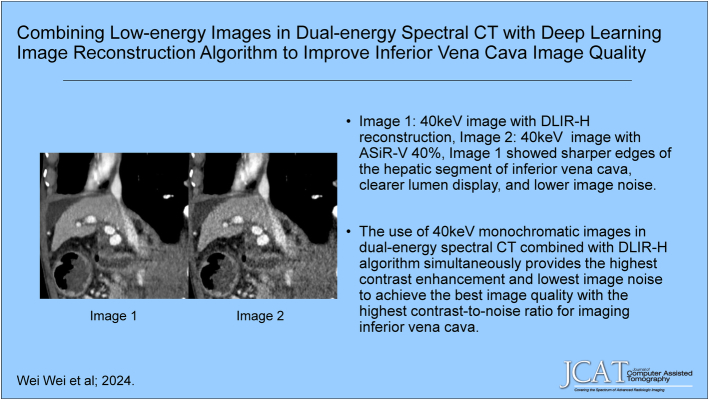
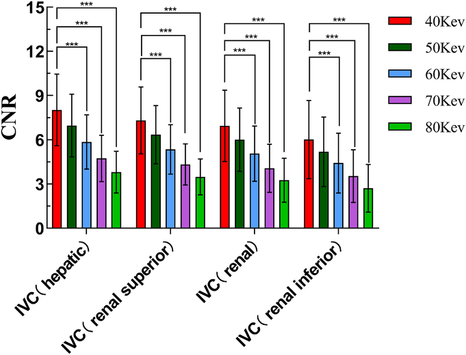
Results: The objective CT value, SD, SNR, and CNR increased with the decrease in energy level, with statistically significant differences (all P <0.05). The 40 keV images had the highest CT values, SNR, and CNR and good diagnostic acceptability, and 40 keV was selected as the best energy level. Compared with ASiR-V40% and DLIR-M, DLIR-H had the lowest SD, highest SNR and CNR, and subjective score (all P <0.001) with good consistencies between the 2 physicians (all k ≥0.75). The 40 keV images with DLIR-H had the highest overall image quality, showing sharper edges of inferior vena cava vessels and clearer lumen in patients with Budd-Chiari syndrome.
Conclusions: Compared with the ASiR-V algorithm, DLIR-H significantly reduces image noise and provides the highest CNR and best diagnostic image quality for the 40 keV DEsCT images in imaging inferior vena cava.
期刊介绍:
The mission of Journal of Computer Assisted Tomography is to showcase the latest clinical and research developments in CT, MR, and closely related diagnostic techniques. We encourage submission of both original research and review articles that have immediate or promissory clinical applications. Topics of special interest include: 1) functional MR and CT of the brain and body; 2) advanced/innovative MRI techniques (diffusion, perfusion, rapid scanning); and 3) advanced/innovative CT techniques (perfusion, multi-energy, dose-reduction, and processing).

 求助内容:
求助内容: 应助结果提醒方式:


