Effect of Manipulation Methods and Storage Environments on the Microstructural, Chemical, and Mechanical Properties of Calcium-Enriched Mixture Cement.
{"title":"Effect of Manipulation Methods and Storage Environments on the Microstructural, Chemical, and Mechanical Properties of Calcium-Enriched Mixture Cement.","authors":"Leyla Roghanizadeh, Hassan Torabzadeh, Ardavan Parhizkar, Alireza Akbarzadeh Baghban, Saeed Asgary","doi":"10.1155/ijbm/5560351","DOIUrl":null,"url":null,"abstract":"<p><p>This study aimed to evaluate the impact of different manipulation methods and storage environments on the microstructural, chemical, and mechanical properties of calcium-enriched mixture (CEM) cement. Four sample groups were examined, including nondried (ND-I) and dried (D-I) groups placed directly in an incubator, dried samples stored in phosphate-buffered saline (PBS) (D-P), and dried samples stored in distilled water (D-W). Various analyses, including Vickers microhardness, compressive strength, Fourier transform infrared spectroscopy (FTIR), X-ray diffraction (XRD), and scanning electron microscopy (SEM) with energy-dispersive X-ray spectroscopy (EDS) were conducted after incubating the samples for 7 days. The data were analyzed by Shapiro-Wilk, Levene, independent <i>t</i>, one-way ANOVA, and Tukey HSD tests. Key findings include the ND-I group exhibited a significantly longer setting time but the lowest microhardness and compressive strength. D-P showed the highest microhardness, while D-W displayed the highest compressive strength. FTIR analysis revealed vibration modes related to (PO4)<sup>3-</sup> ions and Si compounds in all groups, with dried groups showing more vibrations of (PO4)<sup>3-</sup> ions and OH groups, and D-P and D-W groups displayed vibration modes of (CO3)<sup>2-</sup> ions. XRD analysis indicated increased tri/dicalcium silicate reflections in CEM groups exposed to PBS or distilled water. D-I and D-W groups presented hexagonal or rectangular cubic and needle-like crystals, while D-P showed a homogeneous globular structure covered with fine crystals. The order of the weight percentage of major elemental constituents of D-P group was oxygen, calcium, phosphorus, zirconium, barium, carbon, silicon, and sulfur. Incremental placement, drying each increment, and exposing CEM to PBS/tissue fluids result in a faster set and more tolerant cement with a more uniform microstructure. The formation of hydroxyapatite can occur on the surface of the set cement.</p>","PeriodicalId":13704,"journal":{"name":"International Journal of Biomaterials","volume":"2025 ","pages":"5560351"},"PeriodicalIF":4.5000,"publicationDate":"2025-01-20","publicationTypes":"Journal Article","fieldsOfStudy":null,"isOpenAccess":false,"openAccessPdf":"https://www.ncbi.nlm.nih.gov/pmc/articles/PMC11772065/pdf/","citationCount":"0","resultStr":null,"platform":"Semanticscholar","paperid":null,"PeriodicalName":"International Journal of Biomaterials","FirstCategoryId":"1085","ListUrlMain":"https://doi.org/10.1155/ijbm/5560351","RegionNum":0,"RegionCategory":null,"ArticlePicture":[],"TitleCN":null,"AbstractTextCN":null,"PMCID":null,"EPubDate":"2025/1/1 0:00:00","PubModel":"eCollection","JCR":"Q3","JCRName":"MATERIALS SCIENCE, BIOMATERIALS","Score":null,"Total":0}
引用次数: 0
Abstract
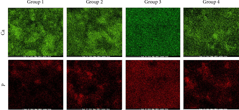
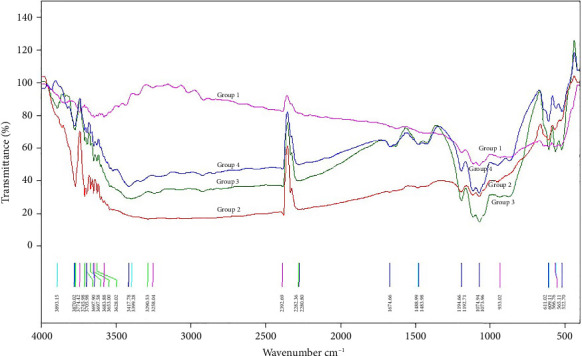
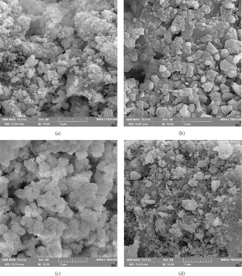
This study aimed to evaluate the impact of different manipulation methods and storage environments on the microstructural, chemical, and mechanical properties of calcium-enriched mixture (CEM) cement. Four sample groups were examined, including nondried (ND-I) and dried (D-I) groups placed directly in an incubator, dried samples stored in phosphate-buffered saline (PBS) (D-P), and dried samples stored in distilled water (D-W). Various analyses, including Vickers microhardness, compressive strength, Fourier transform infrared spectroscopy (FTIR), X-ray diffraction (XRD), and scanning electron microscopy (SEM) with energy-dispersive X-ray spectroscopy (EDS) were conducted after incubating the samples for 7 days. The data were analyzed by Shapiro-Wilk, Levene, independent t, one-way ANOVA, and Tukey HSD tests. Key findings include the ND-I group exhibited a significantly longer setting time but the lowest microhardness and compressive strength. D-P showed the highest microhardness, while D-W displayed the highest compressive strength. FTIR analysis revealed vibration modes related to (PO4)3- ions and Si compounds in all groups, with dried groups showing more vibrations of (PO4)3- ions and OH groups, and D-P and D-W groups displayed vibration modes of (CO3)2- ions. XRD analysis indicated increased tri/dicalcium silicate reflections in CEM groups exposed to PBS or distilled water. D-I and D-W groups presented hexagonal or rectangular cubic and needle-like crystals, while D-P showed a homogeneous globular structure covered with fine crystals. The order of the weight percentage of major elemental constituents of D-P group was oxygen, calcium, phosphorus, zirconium, barium, carbon, silicon, and sulfur. Incremental placement, drying each increment, and exposing CEM to PBS/tissue fluids result in a faster set and more tolerant cement with a more uniform microstructure. The formation of hydroxyapatite can occur on the surface of the set cement.

 求助内容:
求助内容: 应助结果提醒方式:


