Hayrunisa Kahraman Esen, Burcu Biltekin, Mevlit Korkmaz, B Haluk Güvenç
{"title":"Population Kinetics and Protein Profiles of Co-Cultured Adult and Fetus Rabbit Bladder Smooth Muscle Cells.","authors":"Hayrunisa Kahraman Esen, Burcu Biltekin, Mevlit Korkmaz, B Haluk Güvenç","doi":"10.5152/tud.2025.24120","DOIUrl":null,"url":null,"abstract":"<p><strong>Objective: </strong>Bladder tissue models have been developed using smooth muscle cells (SMCs) on various scaffolds to mimic bladder morphology and physiology. This study investigates the effects of co-culturing fetal and adult SMCs on growth properties and protein profiles to understand cellular interactions and population kinetics.</p><p><strong>Methods: </strong>Bladder tissue samples from 10 adult and 10 fetal New Zealand rabbits were divided into 5 groups: adult SMCs (A), fetal SMCs (F), 50%A+50%F (A+F), 75%A+25%F (3A+F), and 25%A+75%F (A+3F). Population doubling time (PDT) of 3 × 10⁶ cells from each group was measured after 48 and 72 hours. Protein concentrations were estimated by spectrophotometric analysis and analyzed via SDS-PAGE gel electrophoresis. Cells exhibited typical SMC morphology, confirmed by positive staining for α-SMA and MYH11.</p><p><strong>Results: </strong>Median cell counts of single cultures were significantly higher than co-cultures (P < .05), but cell viability was comparable (P > .05). Population doubling time at 72 hours for A, F, A+F, 3A+F, and A+3F were 89.4, 92.0, 89.4, 127.9, and 145.0 hours, respectively. Protein concentrations were similar between fetal and adult co-cultures (P > .05). Electrophoresis at 48 hours revealed a unique 80kDa band in adult cells and a 32kDa band in co-cultured cells.</p><p><strong>Conclusion: </strong>Co-culturing resulted in increased PDT, altered protein concentrations, and changes in protein profiles, while each cell population maintained its phenotype. Fetal bladder SMCs maintained their morphology and viability when co-cultured with adult SMCs, resulting in a significant limitation in the cumulative proliferation rate. This may be dependent on alterations of protein profiles of adult and fetal SMCs promoted by rearrangements in co-cultures.</p>","PeriodicalId":101337,"journal":{"name":"Urology research & practice","volume":"50 4","pages":"240-246"},"PeriodicalIF":1.1000,"publicationDate":"2025-01-03","publicationTypes":"Journal Article","fieldsOfStudy":null,"isOpenAccess":false,"openAccessPdf":"https://www.ncbi.nlm.nih.gov/pmc/articles/PMC11883674/pdf/","citationCount":"0","resultStr":null,"platform":"Semanticscholar","paperid":null,"PeriodicalName":"Urology research & practice","FirstCategoryId":"1085","ListUrlMain":"https://doi.org/10.5152/tud.2025.24120","RegionNum":0,"RegionCategory":null,"ArticlePicture":[],"TitleCN":null,"AbstractTextCN":null,"PMCID":null,"EPubDate":"","PubModel":"","JCR":"0","JCRName":"UROLOGY & NEPHROLOGY","Score":null,"Total":0}
引用次数: 0
Abstract
Objective: Bladder tissue models have been developed using smooth muscle cells (SMCs) on various scaffolds to mimic bladder morphology and physiology. This study investigates the effects of co-culturing fetal and adult SMCs on growth properties and protein profiles to understand cellular interactions and population kinetics.
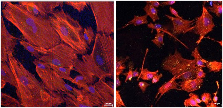
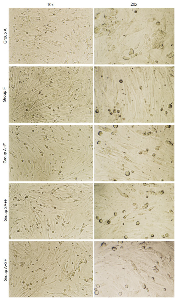
Methods: Bladder tissue samples from 10 adult and 10 fetal New Zealand rabbits were divided into 5 groups: adult SMCs (A), fetal SMCs (F), 50%A+50%F (A+F), 75%A+25%F (3A+F), and 25%A+75%F (A+3F). Population doubling time (PDT) of 3 × 10⁶ cells from each group was measured after 48 and 72 hours. Protein concentrations were estimated by spectrophotometric analysis and analyzed via SDS-PAGE gel electrophoresis. Cells exhibited typical SMC morphology, confirmed by positive staining for α-SMA and MYH11.
Results: Median cell counts of single cultures were significantly higher than co-cultures (P < .05), but cell viability was comparable (P > .05). Population doubling time at 72 hours for A, F, A+F, 3A+F, and A+3F were 89.4, 92.0, 89.4, 127.9, and 145.0 hours, respectively. Protein concentrations were similar between fetal and adult co-cultures (P > .05). Electrophoresis at 48 hours revealed a unique 80kDa band in adult cells and a 32kDa band in co-cultured cells.
Conclusion: Co-culturing resulted in increased PDT, altered protein concentrations, and changes in protein profiles, while each cell population maintained its phenotype. Fetal bladder SMCs maintained their morphology and viability when co-cultured with adult SMCs, resulting in a significant limitation in the cumulative proliferation rate. This may be dependent on alterations of protein profiles of adult and fetal SMCs promoted by rearrangements in co-cultures.


 求助内容:
求助内容: 应助结果提醒方式:


