{"title":"Decoding the molecular enigma behind asbestos and fibrous nanomaterial-induced carcinogenesis.","authors":"Shinya Toyokuni, Yingyi Kong","doi":"10.1093/joccuh/uiae064","DOIUrl":null,"url":null,"abstract":"<p><strong>Objectives: </strong>The natural fibrous mineral, asbestos, has been useful in industry for many centuries. In the 1960s, epidemiology recognized the association between asbestos exposure and mesothelioma, and in 1987 the International Agency for Research on Cancer designated all kinds of asbestos as Group 1 carcinogens. However, various scientific enigmas remained regarding the molecular mechanisms of asbestos-induced mesothelial carcinogenesis. This review article was undertaken to reveal and summarize recent discoveries to resolve those enigmas.</p><p><strong>Methods: </strong>We collected recent important findings from our own laboratory and others to explain why mesothelial cells are the target for asbestos-induced carcinogenesis and what are the key molecular mechanisms.</p><p><strong>Results: </strong>The long incubation period of 30-40 years for mesothelial carcinogenesis after asbestos exposure allows the asbestos fibers to go through the pulmonary parenchyma from the central to peripheral portions and ultimately reach the parietal mesothelium by piercing visceral pleura. Asbestos fibers have affinity for hemoglobin and histones, thus accumulating iron on the surface while traveling through the lung. Mesothelial cells are phagocytic cells, engulfing iron-coated asbestos fibers. Accordingly, homozygous deletion of the p16INK4a tumor suppressor gene, a signature of excess iron-induced carcinogenesis, is acquired through oxidative DNA damage. Recently, exosome-dependent iron transfer from asbestos-fed macrophages to mesothelial cells was reported. Similar molecular mechanisms are observed with multiwalled carbon nanotubes of ~50-nm diameter.</p><p><strong>Conclusions: </strong>Physical dimensions, biopersistence, and affinity to iron/histones are essential for fibrous material to be carcinogenic to mesothelial cells. Therefore, local iron reduction may be a strategy to prevent mesothelial carcinogenesis.</p>","PeriodicalId":16632,"journal":{"name":"Journal of Occupational Health","volume":" ","pages":""},"PeriodicalIF":2.0000,"publicationDate":"2025-01-07","publicationTypes":"Journal Article","fieldsOfStudy":null,"isOpenAccess":false,"openAccessPdf":"https://www.ncbi.nlm.nih.gov/pmc/articles/PMC11830428/pdf/","citationCount":"0","resultStr":null,"platform":"Semanticscholar","paperid":null,"PeriodicalName":"Journal of Occupational Health","FirstCategoryId":"3","ListUrlMain":"https://doi.org/10.1093/joccuh/uiae064","RegionNum":4,"RegionCategory":"医学","ArticlePicture":[],"TitleCN":null,"AbstractTextCN":null,"PMCID":null,"EPubDate":"","PubModel":"","JCR":"Q2","JCRName":"PUBLIC, ENVIRONMENTAL & OCCUPATIONAL HEALTH","Score":null,"Total":0}
引用次数: 0
Abstract
Objectives: The natural fibrous mineral, asbestos, has been useful in industry for many centuries. In the 1960s, epidemiology recognized the association between asbestos exposure and mesothelioma, and in 1987 the International Agency for Research on Cancer designated all kinds of asbestos as Group 1 carcinogens. However, various scientific enigmas remained regarding the molecular mechanisms of asbestos-induced mesothelial carcinogenesis. This review article was undertaken to reveal and summarize recent discoveries to resolve those enigmas.
Methods: We collected recent important findings from our own laboratory and others to explain why mesothelial cells are the target for asbestos-induced carcinogenesis and what are the key molecular mechanisms.
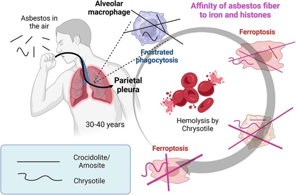
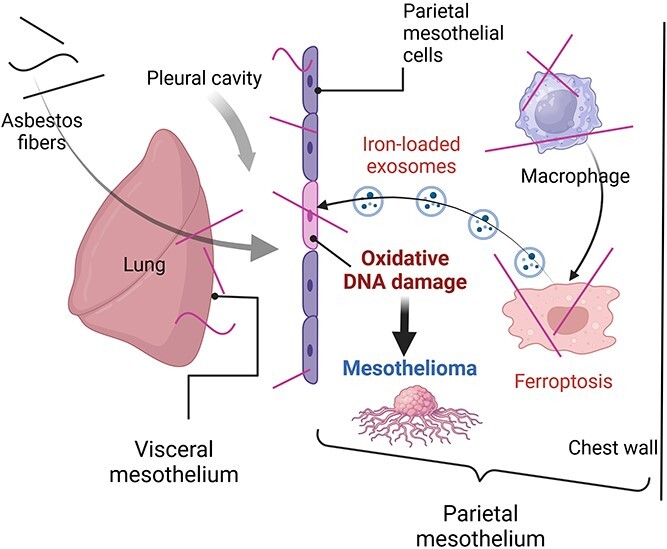
Results: The long incubation period of 30-40 years for mesothelial carcinogenesis after asbestos exposure allows the asbestos fibers to go through the pulmonary parenchyma from the central to peripheral portions and ultimately reach the parietal mesothelium by piercing visceral pleura. Asbestos fibers have affinity for hemoglobin and histones, thus accumulating iron on the surface while traveling through the lung. Mesothelial cells are phagocytic cells, engulfing iron-coated asbestos fibers. Accordingly, homozygous deletion of the p16INK4a tumor suppressor gene, a signature of excess iron-induced carcinogenesis, is acquired through oxidative DNA damage. Recently, exosome-dependent iron transfer from asbestos-fed macrophages to mesothelial cells was reported. Similar molecular mechanisms are observed with multiwalled carbon nanotubes of ~50-nm diameter.
Conclusions: Physical dimensions, biopersistence, and affinity to iron/histones are essential for fibrous material to be carcinogenic to mesothelial cells. Therefore, local iron reduction may be a strategy to prevent mesothelial carcinogenesis.
期刊介绍:
The scope of the journal is broad, covering toxicology, ergonomics, psychosocial factors and other relevant health issues of workers, with special emphasis on the current developments in occupational health. The JOH also accepts various methodologies that are relevant to investigation of occupational health risk factors and exposures, such as large-scale epidemiological studies, human studies employing biological techniques and fundamental experiments on animals, and also welcomes submissions concerning occupational health practices and related issues.

 求助内容:
求助内容: 应助结果提醒方式:


