Raju Jaiswal, Aldina Pivodic, Michail Zoulakis, Kristian F Axelsson, Henrik Litsne, Lisa Johansson, Mattias Lorentzon
{"title":"Prediction of hip fracture by high-resolution peripheral quantitative computed tomography in older Swedish women.","authors":"Raju Jaiswal, Aldina Pivodic, Michail Zoulakis, Kristian F Axelsson, Henrik Litsne, Lisa Johansson, Mattias Lorentzon","doi":"10.1093/jbmr/zjaf020","DOIUrl":null,"url":null,"abstract":"<p><p>The socioeconomic burden of hip fractures, the most severe osteoporotic fracture outcome, is increasing and the current clinical risk assessment lacks sensitivity. This study aimed to develop a method for improved prediction of hip fracture by incorporating measurements of bone microstructure and composition derived from HR-pQCT. In a prospective cohort study of 3028 community-dwelling women aged 75-80, all participants answered questionnaires and underwent baseline examinations of anthropometrics and bone by DXA and HR-pQCT. Medical records, a regional x-ray archive, and registers were used to identify incident fractures and death. Prediction models for hip, major osteoporotic fracture (MOF), and any fracture were developed using Cox proportional hazards regression and machine learning algorithms (neural network, random forest, ensemble, and Extreme Gradient Boosting). In the 2856 (94.3%) women with complete HR-pQCT data at 2 tibia sites (distal and ultra-distal), the median follow-up period was 8.0 yr, and 217 hip, 746 MOF, and 1008 any type of incident fracture occurred. In Cox regression models adjusted for age, BMI, clinical risk factors (CRFs), and FN BMD, the strongest predictors of hip fracture were tibia total volumetric BMD and cortical thickness. The performance of the Cox regression-based prediction models for hip fracture was significantly improved by HR-pQCT (time-dependent AUC; area under receiver operating characteristic curve at 5 yr of follow-up 0.75 [0.64-0.85]), compared to a reference model including CRFs and FN BMD (AUC = 0.71 [0.58-0.81], p < .001) and a Fracture Risk Assessment Tool risk score model (AUC = 0.70 [0.60-0.80], p < .001). The Cox regression model for hip fracture had a significantly higher accuracy than the neural network-based model, the best-performing machine learning algorithm, at clinically relevant sensitivity levels. We conclude that the addition of HR-pQCT parameters improves the prediction of hip fractures in a cohort of older Swedish women.</p>","PeriodicalId":185,"journal":{"name":"Journal of Bone and Mineral Research","volume":" ","pages":"779-790"},"PeriodicalIF":5.9000,"publicationDate":"2025-06-03","publicationTypes":"Journal Article","fieldsOfStudy":null,"isOpenAccess":false,"openAccessPdf":"https://www.ncbi.nlm.nih.gov/pmc/articles/PMC12131241/pdf/","citationCount":"0","resultStr":null,"platform":"Semanticscholar","paperid":null,"PeriodicalName":"Journal of Bone and Mineral Research","FirstCategoryId":"3","ListUrlMain":"https://doi.org/10.1093/jbmr/zjaf020","RegionNum":1,"RegionCategory":"医学","ArticlePicture":[],"TitleCN":null,"AbstractTextCN":null,"PMCID":null,"EPubDate":"","PubModel":"","JCR":"Q1","JCRName":"ENDOCRINOLOGY & METABOLISM","Score":null,"Total":0}
引用次数: 0
Abstract
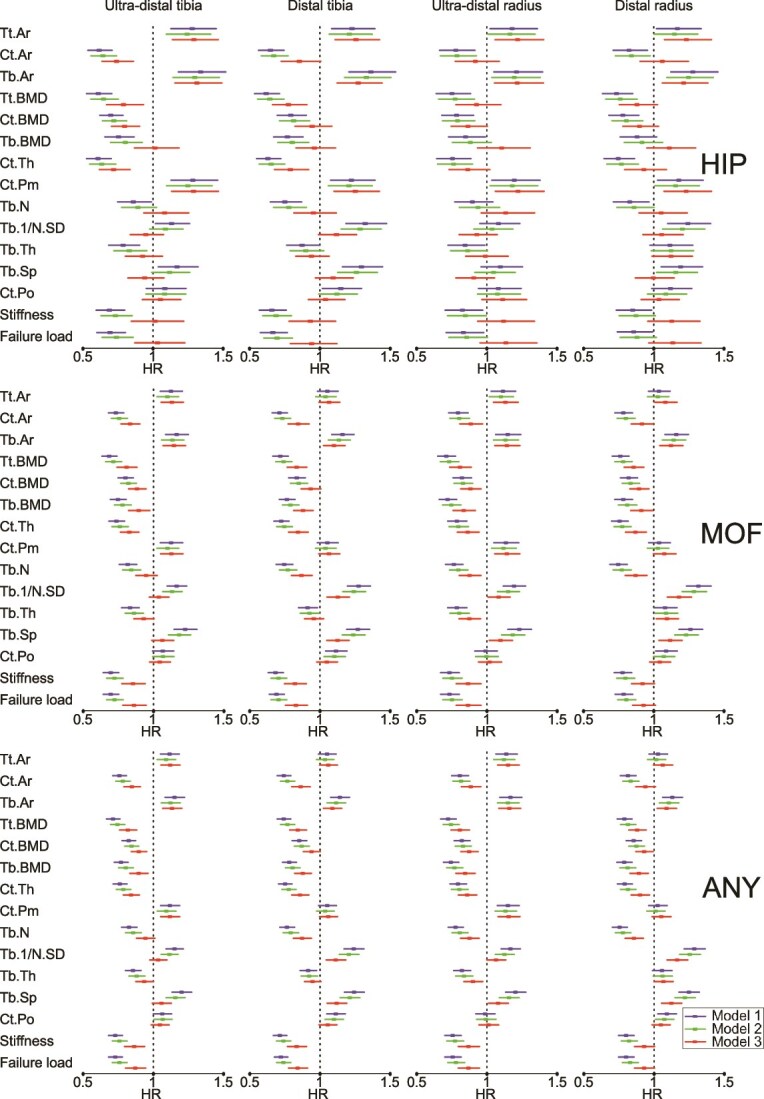
The socioeconomic burden of hip fractures, the most severe osteoporotic fracture outcome, is increasing and the current clinical risk assessment lacks sensitivity. This study aimed to develop a method for improved prediction of hip fracture by incorporating measurements of bone microstructure and composition derived from HR-pQCT. In a prospective cohort study of 3028 community-dwelling women aged 75-80, all participants answered questionnaires and underwent baseline examinations of anthropometrics and bone by DXA and HR-pQCT. Medical records, a regional x-ray archive, and registers were used to identify incident fractures and death. Prediction models for hip, major osteoporotic fracture (MOF), and any fracture were developed using Cox proportional hazards regression and machine learning algorithms (neural network, random forest, ensemble, and Extreme Gradient Boosting). In the 2856 (94.3%) women with complete HR-pQCT data at 2 tibia sites (distal and ultra-distal), the median follow-up period was 8.0 yr, and 217 hip, 746 MOF, and 1008 any type of incident fracture occurred. In Cox regression models adjusted for age, BMI, clinical risk factors (CRFs), and FN BMD, the strongest predictors of hip fracture were tibia total volumetric BMD and cortical thickness. The performance of the Cox regression-based prediction models for hip fracture was significantly improved by HR-pQCT (time-dependent AUC; area under receiver operating characteristic curve at 5 yr of follow-up 0.75 [0.64-0.85]), compared to a reference model including CRFs and FN BMD (AUC = 0.71 [0.58-0.81], p < .001) and a Fracture Risk Assessment Tool risk score model (AUC = 0.70 [0.60-0.80], p < .001). The Cox regression model for hip fracture had a significantly higher accuracy than the neural network-based model, the best-performing machine learning algorithm, at clinically relevant sensitivity levels. We conclude that the addition of HR-pQCT parameters improves the prediction of hip fractures in a cohort of older Swedish women.
期刊介绍:
The Journal of Bone and Mineral Research (JBMR) publishes highly impactful original manuscripts, reviews, and special articles on basic, translational and clinical investigations relevant to the musculoskeletal system and mineral metabolism. Specifically, the journal is interested in original research on the biology and physiology of skeletal tissues, interdisciplinary research spanning the musculoskeletal and other systems, including but not limited to immunology, hematology, energy metabolism, cancer biology, and neurology, and systems biology topics using large scale “-omics” approaches. The journal welcomes clinical research on the pathophysiology, treatment and prevention of osteoporosis and fractures, as well as sarcopenia, disorders of bone and mineral metabolism, and rare or genetically determined bone diseases.
 求助内容:
求助内容: 应助结果提醒方式:


