The interaction between third molars and surrounding periapical tissues in mandibular stress distribution during high-impact trauma: a finite element study.
{"title":"The interaction between third molars and surrounding periapical tissues in mandibular stress distribution during high-impact trauma: a finite element study.","authors":"C-B Nogueira, F-W Costa, F-S Carvalho, T-P Bezerra, I-C Neto, F-I Júnior, E-C Soares","doi":"10.4317/medoral.26954","DOIUrl":null,"url":null,"abstract":"<p><strong>Background: </strong>The presence of mandibular third molars has been associated with the risk of mandibular fractures, highlighting the need for comprehensive studies considering the interaction with other mandibular structures. This study investigates how mandibular third molars and neighboring tissues can influence the structural fragility of the mandible using finite element analysis.</p><p><strong>Material and methods: </strong>A finite element analysis study following the guidelines proposed by RIFEM 1.0 was performed using three previously created mandible models: Model A, without right and left third molars; Model B, without one third molar; Model C, with bilateral presence of third molars. A 2452N force was applied to the right mandibular body in a virtual environment, allowing for a structural analysis of each mandible.</p><p><strong>Results: </strong>Models without third molars and with only one third molar showed similar energy dissipation patterns, contrasting with the model with both third molars. The presence of third molars influenced the magnitude and distribution of stress, highlighting fragility points in specific areas such as the lingual surface, the condyles bilaterally (models without and with one contralateral third molar to trauma), and the distal cervical region of the second molar (third molar absent), as well as significantly showed the path of energy towards the contralateral side of the trauma with a concentration of energy at the contact points of virtually all teeth present immediately after impact.</p><p><strong>Conclusions: </strong>The presence of mandibular third molars influenced the distribution and magnitude of stress within the mandible during a simulated high-impact trauma. Models with third molars exhibit distinct stress patterns, with fragility points appearing in critical areas such as the lingual surface, condyles, and second molar regions. These findings suggest that the presence of third molars increases the structural fragility of the mandible, potentially elevating the risk of mandibular fractures, especially in the context of traumatic impacts.</p>","PeriodicalId":49016,"journal":{"name":"Medicina Oral Patologia Oral Y Cirugia Bucal","volume":" ","pages":"e394-e400"},"PeriodicalIF":2.1000,"publicationDate":"2025-05-01","publicationTypes":"Journal Article","fieldsOfStudy":null,"isOpenAccess":false,"openAccessPdf":"https://www.ncbi.nlm.nih.gov/pmc/articles/PMC12019648/pdf/","citationCount":"0","resultStr":null,"platform":"Semanticscholar","paperid":null,"PeriodicalName":"Medicina Oral Patologia Oral Y Cirugia Bucal","FirstCategoryId":"3","ListUrlMain":"https://doi.org/10.4317/medoral.26954","RegionNum":3,"RegionCategory":"医学","ArticlePicture":[],"TitleCN":null,"AbstractTextCN":null,"PMCID":null,"EPubDate":"","PubModel":"","JCR":"Q2","JCRName":"DENTISTRY, ORAL SURGERY & MEDICINE","Score":null,"Total":0}
引用次数: 0
Abstract
Background: The presence of mandibular third molars has been associated with the risk of mandibular fractures, highlighting the need for comprehensive studies considering the interaction with other mandibular structures. This study investigates how mandibular third molars and neighboring tissues can influence the structural fragility of the mandible using finite element analysis.
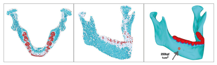
Material and methods: A finite element analysis study following the guidelines proposed by RIFEM 1.0 was performed using three previously created mandible models: Model A, without right and left third molars; Model B, without one third molar; Model C, with bilateral presence of third molars. A 2452N force was applied to the right mandibular body in a virtual environment, allowing for a structural analysis of each mandible.
Results: Models without third molars and with only one third molar showed similar energy dissipation patterns, contrasting with the model with both third molars. The presence of third molars influenced the magnitude and distribution of stress, highlighting fragility points in specific areas such as the lingual surface, the condyles bilaterally (models without and with one contralateral third molar to trauma), and the distal cervical region of the second molar (third molar absent), as well as significantly showed the path of energy towards the contralateral side of the trauma with a concentration of energy at the contact points of virtually all teeth present immediately after impact.
Conclusions: The presence of mandibular third molars influenced the distribution and magnitude of stress within the mandible during a simulated high-impact trauma. Models with third molars exhibit distinct stress patterns, with fragility points appearing in critical areas such as the lingual surface, condyles, and second molar regions. These findings suggest that the presence of third molars increases the structural fragility of the mandible, potentially elevating the risk of mandibular fractures, especially in the context of traumatic impacts.
期刊介绍:
1. Oral Medicine and Pathology:
Clinicopathological as well as medical or surgical management aspects of
diseases affecting oral mucosa, salivary glands, maxillary bones, as well as
orofacial neurological disorders, and systemic conditions with an impact on
the oral cavity.
2. Oral Surgery:
Surgical management aspects of diseases affecting oral mucosa, salivary glands,
maxillary bones, teeth, implants, oral surgical procedures. Surgical management
of diseases affecting head and neck areas.
3. Medically compromised patients in Dentistry:
Articles discussing medical problems in Odontology will also be included, with
a special focus on the clinico-odontological management of medically compromised patients, and considerations regarding high-risk or disabled patients.
4. Implantology
5. Periodontology

 求助内容:
求助内容: 应助结果提醒方式:


