Ziwei Wang, Jicheng Xiong, Lin Peng, Xiaobo Wu, Yongtao Han, Yi Zhu, Xuefeng Leng
{"title":"Giant thymolipoma in a 16-year-old girl with multimodal diagnostic approach and surgical management: a case report.","authors":"Ziwei Wang, Jicheng Xiong, Lin Peng, Xiaobo Wu, Yongtao Han, Yi Zhu, Xuefeng Leng","doi":"10.21037/acr-24-157","DOIUrl":null,"url":null,"abstract":"<p><strong>Background: </strong>Thymolipomas are rare benign mediastinal tumors primarily occurring in young adults, although they can also present in pediatric populations. These tumors are often asymptomatic, but their substantial size can create significant diagnostic and therapeutic challenges, necessitating careful evaluation and management.</p><p><strong>Case description: </strong>A teenage girl was diagnosed with a giant thymolipoma, which was discovered incidentally during a routine chest radiograph. Notably, the patient remained asymptomatic despite the tumor's considerable size and its apparent impact on surrounding thoracic structures. To facilitate a thorough preoperative assessment, a multimodal imaging approach was employed, including contrast-enhanced ultrasound (CEUS), computed tomography (CT), and magnetic resonance imaging (MRI). These advanced imaging techniques played a crucial role in delineating the tumor's extent, characteristics, and relationship to adjacent anatomical structures, thereby informing surgical planning. Ultimately, the tumor was successfully excised through a median sternotomy. Postoperative pathological examination confirmed the diagnosis of thymolipoma. Remarkably, after a follow-up period of five years, the patient showed no signs of recurrence and maintained a healthy status.</p><p><strong>Conclusions: </strong>This case underscores the effectiveness of a multimodal imaging strategy for the diagnosis and preoperative assessment of pediatric thymolipomas. It emphasizes the feasibility of complete surgical resection, even for large tumors, leading to a favorable prognosis. Moreover, it highlights the importance of tailored management strategies for pediatric patients with rare thoracic tumors, as evidenced by this successful clinical outcome.</p>","PeriodicalId":29752,"journal":{"name":"AME Case Reports","volume":"9 ","pages":"25"},"PeriodicalIF":0.7000,"publicationDate":"2025-01-06","publicationTypes":"Journal Article","fieldsOfStudy":null,"isOpenAccess":false,"openAccessPdf":"https://www.ncbi.nlm.nih.gov/pmc/articles/PMC11759932/pdf/","citationCount":"0","resultStr":null,"platform":"Semanticscholar","paperid":null,"PeriodicalName":"AME Case Reports","FirstCategoryId":"1085","ListUrlMain":"https://doi.org/10.21037/acr-24-157","RegionNum":0,"RegionCategory":null,"ArticlePicture":[],"TitleCN":null,"AbstractTextCN":null,"PMCID":null,"EPubDate":"2025/1/1 0:00:00","PubModel":"eCollection","JCR":"Q3","JCRName":"MEDICINE, GENERAL & INTERNAL","Score":null,"Total":0}
引用次数: 0
Abstract
Background: Thymolipomas are rare benign mediastinal tumors primarily occurring in young adults, although they can also present in pediatric populations. These tumors are often asymptomatic, but their substantial size can create significant diagnostic and therapeutic challenges, necessitating careful evaluation and management.
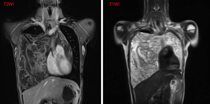
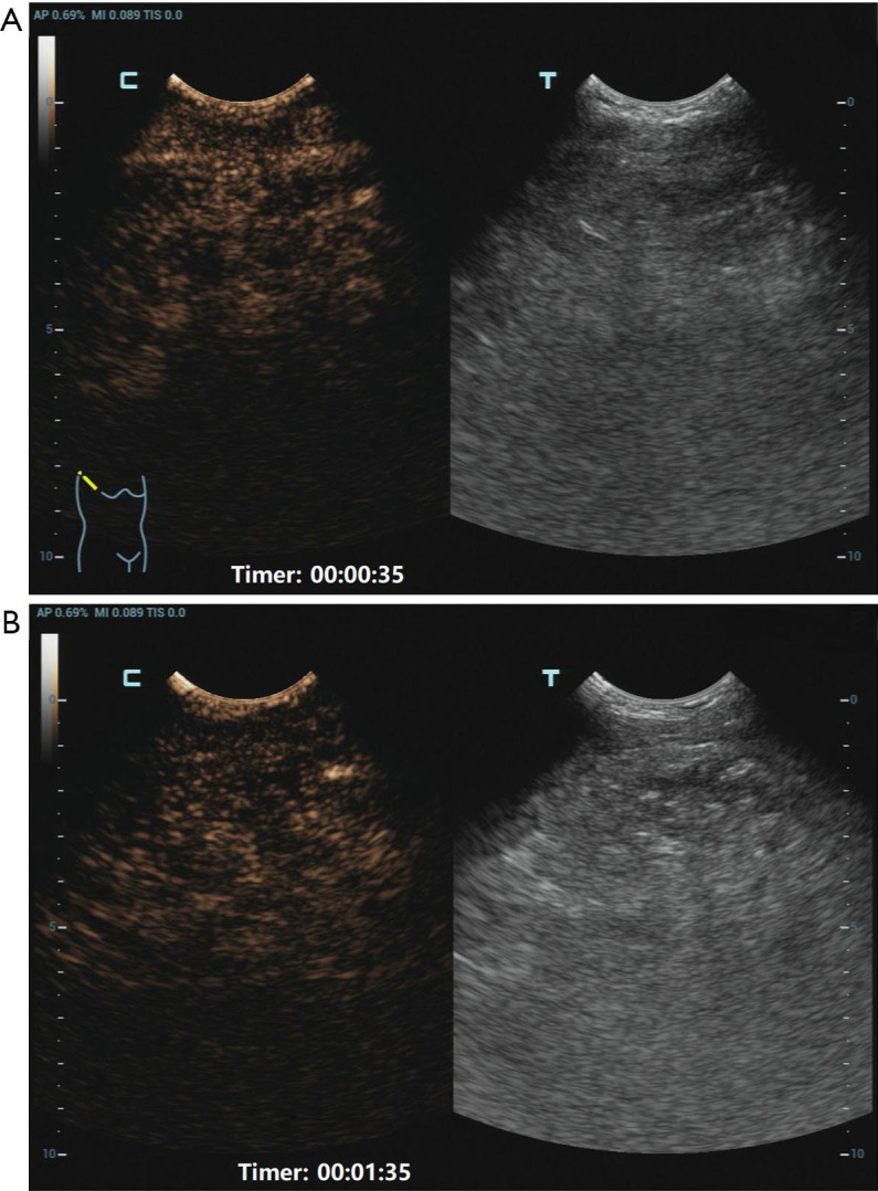
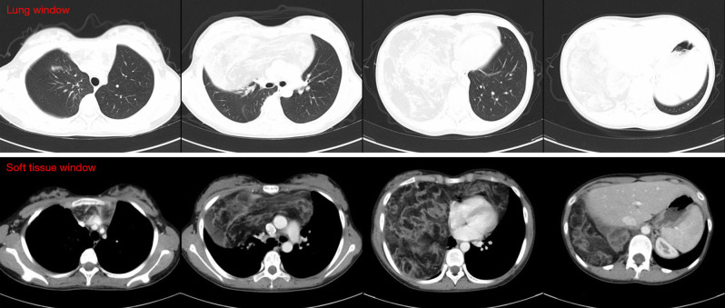
Case description: A teenage girl was diagnosed with a giant thymolipoma, which was discovered incidentally during a routine chest radiograph. Notably, the patient remained asymptomatic despite the tumor's considerable size and its apparent impact on surrounding thoracic structures. To facilitate a thorough preoperative assessment, a multimodal imaging approach was employed, including contrast-enhanced ultrasound (CEUS), computed tomography (CT), and magnetic resonance imaging (MRI). These advanced imaging techniques played a crucial role in delineating the tumor's extent, characteristics, and relationship to adjacent anatomical structures, thereby informing surgical planning. Ultimately, the tumor was successfully excised through a median sternotomy. Postoperative pathological examination confirmed the diagnosis of thymolipoma. Remarkably, after a follow-up period of five years, the patient showed no signs of recurrence and maintained a healthy status.
Conclusions: This case underscores the effectiveness of a multimodal imaging strategy for the diagnosis and preoperative assessment of pediatric thymolipomas. It emphasizes the feasibility of complete surgical resection, even for large tumors, leading to a favorable prognosis. Moreover, it highlights the importance of tailored management strategies for pediatric patients with rare thoracic tumors, as evidenced by this successful clinical outcome.

 求助内容:
求助内容: 应助结果提醒方式:


