Tarcila Gurgel Aquino, Diogenes de Melo Jacó, Ingryd Gabriella Nascimento Santos, Eliauria Rosa Martins
{"title":"Idiopathic pulmonary arteriovenous malformation: a rarity in clinical practice.","authors":"Tarcila Gurgel Aquino, Diogenes de Melo Jacó, Ingryd Gabriella Nascimento Santos, Eliauria Rosa Martins","doi":"10.1590/1677-5449.202400052","DOIUrl":null,"url":null,"abstract":"<p><p>Pulmonary arteriovenous malformations (PAVM) are characterized by abnormal pulmonary vessels forming arteriovenous shunts that compromise oxygenation of the blood, causing hypoxemia, and predispose to infections and cerebral ischemia. The patient in this case was a 38-year-old male who presented with tachypnea and dyspnea, cyanosis of extremities, and significant digital clubbing. The patient had structural epilepsy secondary to neurosurgery for a cerebral abscess during childhood. Arterial blood gas analysis showed significant hypoxemia (PaO<sub>2</sub> = 46.2; SaO<sub>2</sub> = 77%; PaO<sub>2</sub>/FiO<sub>2</sub> = 70) and a chest computed tomography showed PAVM in the apical segments of the right upper and lower lobes, with ectatic and tortuous vascular structures following an intraparenchymal path, communicating with the pulmonary artery and veins. After confirmation of the PAVM, it was concluded that elevated pulmonary resistance was contributing to refractive hypoxemia and hypercapnia. Gradual reduction of the ventilation parameters, primarily controlled pressure and positive end-expiratory pressure, and consequent reduction of the arteriovenous shunt, resulted in progressive improvement of oxygenation and respiratory mechanics. The vascular surgery team's assessment was that treatment with embolization was warranted.</p>","PeriodicalId":14814,"journal":{"name":"Jornal Vascular Brasileiro","volume":"23 ","pages":"e20240005"},"PeriodicalIF":1.0000,"publicationDate":"2025-01-13","publicationTypes":"Journal Article","fieldsOfStudy":null,"isOpenAccess":false,"openAccessPdf":"https://www.ncbi.nlm.nih.gov/pmc/articles/PMC11758929/pdf/","citationCount":"0","resultStr":null,"platform":"Semanticscholar","paperid":null,"PeriodicalName":"Jornal Vascular Brasileiro","FirstCategoryId":"1085","ListUrlMain":"https://doi.org/10.1590/1677-5449.202400052","RegionNum":0,"RegionCategory":null,"ArticlePicture":[],"TitleCN":null,"AbstractTextCN":null,"PMCID":null,"EPubDate":"2024/1/1 0:00:00","PubModel":"eCollection","JCR":"Q4","JCRName":"PERIPHERAL VASCULAR DISEASE","Score":null,"Total":0}
引用次数: 0
Abstract
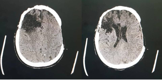
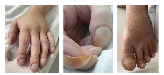
Pulmonary arteriovenous malformations (PAVM) are characterized by abnormal pulmonary vessels forming arteriovenous shunts that compromise oxygenation of the blood, causing hypoxemia, and predispose to infections and cerebral ischemia. The patient in this case was a 38-year-old male who presented with tachypnea and dyspnea, cyanosis of extremities, and significant digital clubbing. The patient had structural epilepsy secondary to neurosurgery for a cerebral abscess during childhood. Arterial blood gas analysis showed significant hypoxemia (PaO2 = 46.2; SaO2 = 77%; PaO2/FiO2 = 70) and a chest computed tomography showed PAVM in the apical segments of the right upper and lower lobes, with ectatic and tortuous vascular structures following an intraparenchymal path, communicating with the pulmonary artery and veins. After confirmation of the PAVM, it was concluded that elevated pulmonary resistance was contributing to refractive hypoxemia and hypercapnia. Gradual reduction of the ventilation parameters, primarily controlled pressure and positive end-expiratory pressure, and consequent reduction of the arteriovenous shunt, resulted in progressive improvement of oxygenation and respiratory mechanics. The vascular surgery team's assessment was that treatment with embolization was warranted.
期刊介绍:
The Jornal Vascular Brasileiro is editated and published quaterly to select and disseminate high-quality scientific contents concerning original research, novel surgical and diagnostic techniques, and clinical observations in the field of vascular surgery, angiology, and endovascular surgery. Its abbreviated title is J. Vasc. Bras., which should be used in bibliographies, footnotes and bibliographical references and strips.


 求助内容:
求助内容: 应助结果提醒方式:


