Enhancing diagnostic accuracy in breast cancer: integrating novel machine learning approaches with enhanced image preprocessing for improved mammography analysis.
{"title":"Enhancing diagnostic accuracy in breast cancer: integrating novel machine learning approaches with enhanced image preprocessing for improved mammography analysis.","authors":"Mohsen Mehrabi, Nafise Salek","doi":"10.5114/pjr/195523","DOIUrl":null,"url":null,"abstract":"<p><strong>Purpose: </strong>This study explored the use of computer-aided diagnosis (CAD) systems to enhance mammography image quality and identify potentially suspicious areas, because mammography is the primary method for breast cancer screening. The primary aim was to find the best combination of preprocessing algorithms to enable more precise classification and interpretation of mammography images because the selected preprocessing algorithms significantly impact the effectiveness of later classification and segmentation processes.</p><p><strong>Material and methods: </strong>The study utilised the mini-MIAS database of mammography images and examined the impact of applying various preprocessing method combinations to differentiate between malignant and benign breast lesions. The preprocessing steps included removing label information and pectoral muscle, followed by applying algorithms such as contrast-limited adaptive histogram equalisation (CLAHE), unsharp masking (USM), and median filtering (MF) to enhance image resolution and visibility. After preprocessing, a <i>k</i>-means clustering technique was used to extract potentially suspicious regions, and features were then extracted from these regions of interest (ROIs). The extracted feature datasets were classified using various machine learning algorithms, including artificial neural networks, random forest, and support vector machines.</p><p><strong>Results: </strong>The findings showed that the combination of CLAHE, USM, and MF preprocessing algorithms resulted in the highest classification performance, outperforming the use of CLAHE alone.</p><p><strong>Conclusions: </strong>The integration of advanced preprocessing techniques with machine learning significantly enhances the accuracy of mammography analysis, facilitating more precise differentiation between malignant and benign breast lesions.</p>","PeriodicalId":94174,"journal":{"name":"Polish journal of radiology","volume":"89 ","pages":"e573-e583"},"PeriodicalIF":0.0000,"publicationDate":"2024-12-22","publicationTypes":"Journal Article","fieldsOfStudy":null,"isOpenAccess":false,"openAccessPdf":"https://www.ncbi.nlm.nih.gov/pmc/articles/PMC11756364/pdf/","citationCount":"0","resultStr":null,"platform":"Semanticscholar","paperid":null,"PeriodicalName":"Polish journal of radiology","FirstCategoryId":"1085","ListUrlMain":"https://doi.org/10.5114/pjr/195523","RegionNum":0,"RegionCategory":null,"ArticlePicture":[],"TitleCN":null,"AbstractTextCN":null,"PMCID":null,"EPubDate":"2024/1/1 0:00:00","PubModel":"eCollection","JCR":"","JCRName":"","Score":null,"Total":0}
引用次数: 0
Abstract
Purpose: This study explored the use of computer-aided diagnosis (CAD) systems to enhance mammography image quality and identify potentially suspicious areas, because mammography is the primary method for breast cancer screening. The primary aim was to find the best combination of preprocessing algorithms to enable more precise classification and interpretation of mammography images because the selected preprocessing algorithms significantly impact the effectiveness of later classification and segmentation processes.
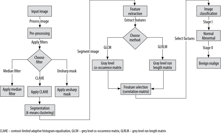
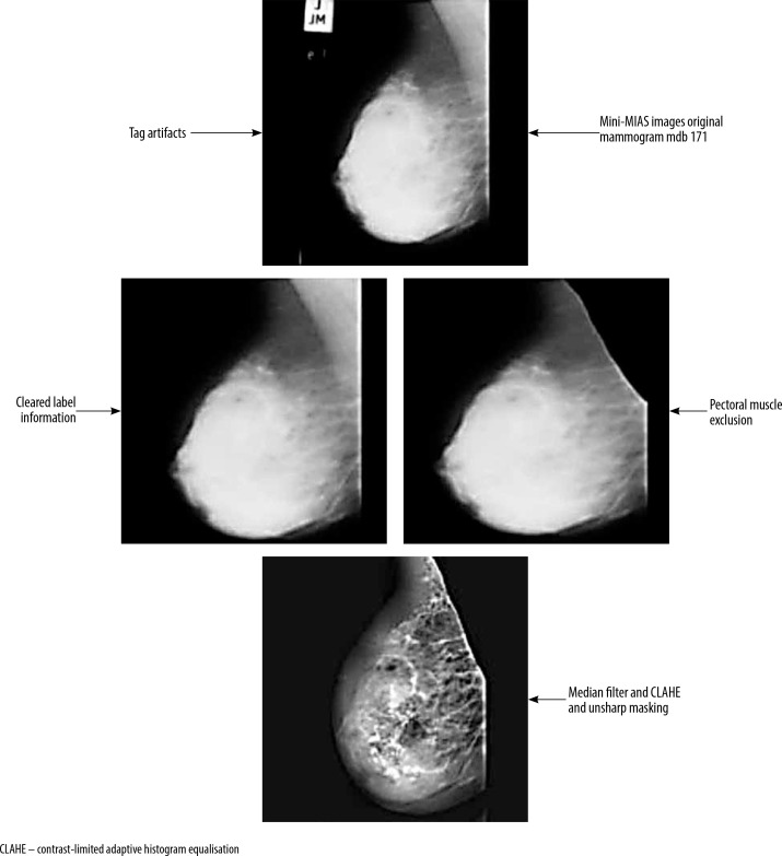
Material and methods: The study utilised the mini-MIAS database of mammography images and examined the impact of applying various preprocessing method combinations to differentiate between malignant and benign breast lesions. The preprocessing steps included removing label information and pectoral muscle, followed by applying algorithms such as contrast-limited adaptive histogram equalisation (CLAHE), unsharp masking (USM), and median filtering (MF) to enhance image resolution and visibility. After preprocessing, a k-means clustering technique was used to extract potentially suspicious regions, and features were then extracted from these regions of interest (ROIs). The extracted feature datasets were classified using various machine learning algorithms, including artificial neural networks, random forest, and support vector machines.
Results: The findings showed that the combination of CLAHE, USM, and MF preprocessing algorithms resulted in the highest classification performance, outperforming the use of CLAHE alone.
Conclusions: The integration of advanced preprocessing techniques with machine learning significantly enhances the accuracy of mammography analysis, facilitating more precise differentiation between malignant and benign breast lesions.


 求助内容:
求助内容: 应助结果提醒方式:


