Seyed Ali Mirshahvalad, Adriano Basso Dias, Claudia Ortega, Jorge Andres Abreu Gomez, Satheesh Krishna, Nathan Perlis, Alejandro Berlin, Theodorus van der Kwast, Kartik Jhaveri, Sangeet Ghai, Ur Metser, Anna Theresa Santiago, Patrick Veit-Haibach
{"title":"[18F]F-DCFPyL PET/MRI radiomics for intraprostatic prostate cancer detection and metastases prediction using whole-gland segmentation.","authors":"Seyed Ali Mirshahvalad, Adriano Basso Dias, Claudia Ortega, Jorge Andres Abreu Gomez, Satheesh Krishna, Nathan Perlis, Alejandro Berlin, Theodorus van der Kwast, Kartik Jhaveri, Sangeet Ghai, Ur Metser, Anna Theresa Santiago, Patrick Veit-Haibach","doi":"10.1093/bjr/tqaf014","DOIUrl":null,"url":null,"abstract":"<p><strong>Objectives: </strong>To evaluate [18F]F-DCFPyL PET/MRI whole-gland-derived radiomics for detecting clinically significant (cs) prostate cancer (PCa) within the prostate gland and predicting extra-prostatic metastasis (N and M staging).</p><p><strong>Methods: </strong>In this single-centre, retrospective study, therapy-naïve PCa patients who underwent [18F]F-DCFPyL PET/MRI were included. Whole-prostate segmentation was performed. Feature extraction from each modality was done. The selection of potential variables was made through regularized binomial logistic regression. The oversampled training data were used to train binomial logistic regression for each outcome. The estimates of the models were calculated, and the mean accuracy was reported. The trained models were assessed on the test data for comparative evaluation of performance.</p><p><strong>Results: </strong>A total of 103 patients (mean age = 65; mean PSA = 23.4) were studied. Among them, 89 had csPCa and 20 had metastatic disease. There were five radiomics variables selected for the International Society of Urological Pathology Grade Group (ISUP GG) ≥ 2 from T2w, ADC, and PET. To detect N1, five radiomics variables were selected from the T2w and PET. For M1, four radiomics variables were selected from T2w and ADC. Regarding the performance of models for the prediction of csPCa, the imaging-based hybrid model (T2w + PET) provided the highest AUC (0.98). The performance of N1 models showed the highest AUC (0.80) for T2w + PET. To predict M1, the T2w + ADC model showed the highest AUC (0.93).</p><p><strong>Conclusions: </strong>Whole-gland PET/MRI radiomics may provide a reliable model to predict csPCa. Also, acceptable performance was reached for predicting metastatic disease in our limited population. Our findings may support the value of whole-gland radiomics for non-invasive csPCa detection and prediction of metastatic disease.</p><p><strong>Advances in knowledge: </strong>Whole-gland PET/MRI radiomics, a less operator-dependent segmentation method, can be potentially used for treatment personalization in PCa patients.</p><p><strong>Trial registration: </strong>NCT03535831. Registered 2018; NCT03149861. Registered 2017.</p>","PeriodicalId":9306,"journal":{"name":"British Journal of Radiology","volume":" ","pages":"1606-1614"},"PeriodicalIF":3.4000,"publicationDate":"2025-10-01","publicationTypes":"Journal Article","fieldsOfStudy":null,"isOpenAccess":false,"openAccessPdf":"https://www.ncbi.nlm.nih.gov/pmc/articles/PMC12515043/pdf/","citationCount":"0","resultStr":null,"platform":"Semanticscholar","paperid":null,"PeriodicalName":"British Journal of Radiology","FirstCategoryId":"3","ListUrlMain":"https://doi.org/10.1093/bjr/tqaf014","RegionNum":4,"RegionCategory":"医学","ArticlePicture":[],"TitleCN":null,"AbstractTextCN":null,"PMCID":null,"EPubDate":"","PubModel":"","JCR":"Q3","JCRName":"RADIOLOGY, NUCLEAR MEDICINE & MEDICAL IMAGING","Score":null,"Total":0}
引用次数: 0
Abstract
Objectives: To evaluate [18F]F-DCFPyL PET/MRI whole-gland-derived radiomics for detecting clinically significant (cs) prostate cancer (PCa) within the prostate gland and predicting extra-prostatic metastasis (N and M staging).
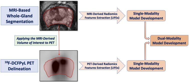
Methods: In this single-centre, retrospective study, therapy-naïve PCa patients who underwent [18F]F-DCFPyL PET/MRI were included. Whole-prostate segmentation was performed. Feature extraction from each modality was done. The selection of potential variables was made through regularized binomial logistic regression. The oversampled training data were used to train binomial logistic regression for each outcome. The estimates of the models were calculated, and the mean accuracy was reported. The trained models were assessed on the test data for comparative evaluation of performance.
Results: A total of 103 patients (mean age = 65; mean PSA = 23.4) were studied. Among them, 89 had csPCa and 20 had metastatic disease. There were five radiomics variables selected for the International Society of Urological Pathology Grade Group (ISUP GG) ≥ 2 from T2w, ADC, and PET. To detect N1, five radiomics variables were selected from the T2w and PET. For M1, four radiomics variables were selected from T2w and ADC. Regarding the performance of models for the prediction of csPCa, the imaging-based hybrid model (T2w + PET) provided the highest AUC (0.98). The performance of N1 models showed the highest AUC (0.80) for T2w + PET. To predict M1, the T2w + ADC model showed the highest AUC (0.93).
Conclusions: Whole-gland PET/MRI radiomics may provide a reliable model to predict csPCa. Also, acceptable performance was reached for predicting metastatic disease in our limited population. Our findings may support the value of whole-gland radiomics for non-invasive csPCa detection and prediction of metastatic disease.
Advances in knowledge: Whole-gland PET/MRI radiomics, a less operator-dependent segmentation method, can be potentially used for treatment personalization in PCa patients.
期刊介绍:
BJR is the international research journal of the British Institute of Radiology and is the oldest scientific journal in the field of radiology and related sciences.
Dating back to 1896, BJR’s history is radiology’s history, and the journal has featured some landmark papers such as the first description of Computed Tomography "Computerized transverse axial tomography" by Godfrey Hounsfield in 1973. A valuable historical resource, the complete BJR archive has been digitized from 1896.
Quick Facts:
- 2015 Impact Factor – 1.840
- Receipt to first decision – average of 6 weeks
- Acceptance to online publication – average of 3 weeks
- ISSN: 0007-1285
- eISSN: 1748-880X
Open Access option

 求助内容:
求助内容: 应助结果提醒方式:


