Lalee Varghese, Rakesh R Bright, Aditya V A Gunturi, Grace Rebekah, Regi Kurien
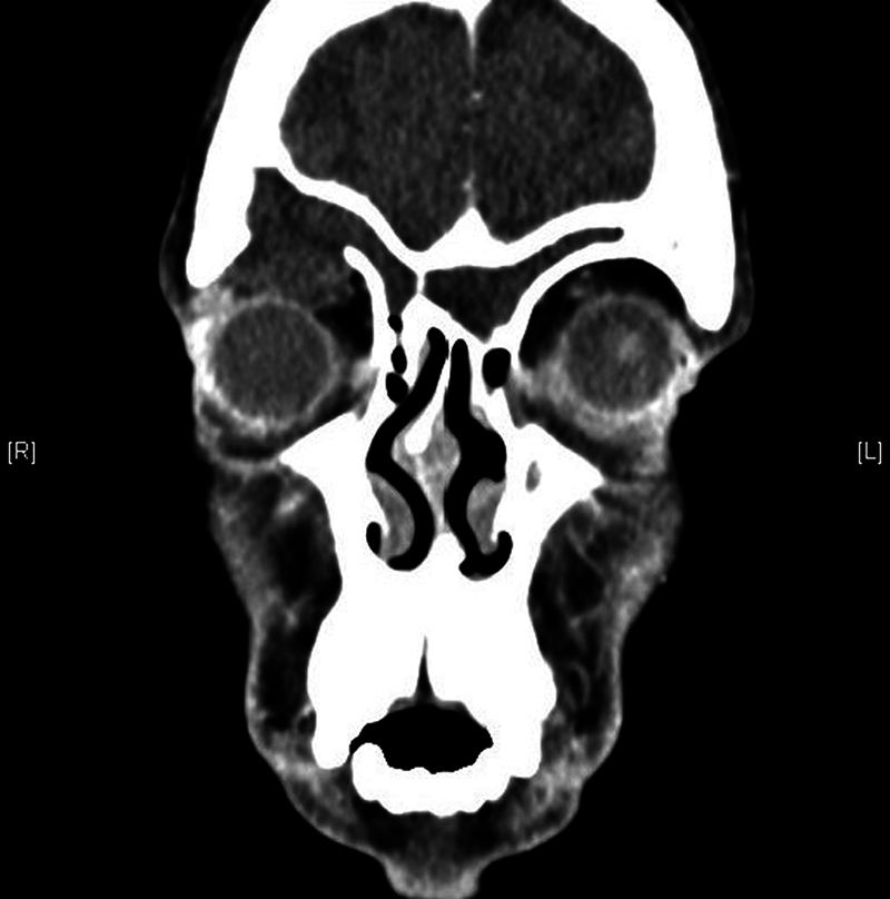
{"title":"Do Variations in Frontal Recess Anatomy Predispose to Mucocele Formation?","authors":"Lalee Varghese, Rakesh R Bright, Aditya V A Gunturi, Grace Rebekah, Regi Kurien","doi":"10.1055/s-0044-1788002","DOIUrl":null,"url":null,"abstract":"<p><p><b>Introduction</b> Mucoceles are benign expansile cystic lesions commonly seen in the frontoethmoidal region. <b>Objective</b> To see if the distribution of frontal air cells predisposes to mucocele formation. <b>Methods</b> Retrospective review of all cases of paranasal sinus mucocele from 2011 to 2021. Data on demographics, history of surgery or trauma, clinical features, radiological findings, and outcome were collected and analyzed. <b>Results</b> Of the 28 cases, 19 (67.9%) were male and 9 (32.1%), female, with a mean age of 40.75 years. Mucocele was unilateral in 26 (92.9%) patients. Twenty patients (71.43%) presented with primary mucocele. The distribution of mucocele was frontal and frontoethmoidal in 8 (28.6%) patients each, maxillary in 6 (21.4%), and ethmoid and sphenoid sinus in 3 (10.7%) patients each. Sixteen (57.1%) patients had frontal sinus involvement. At presentation, 13 (46.4%) patients had nasal symptoms, 17 (60.7%) had orbital symptoms, while 16 (57.1%) had headache. Pain (12; 70.59%) was the predominant orbital symptom, followed by proptosis and diplopia (8; 47.06%). The most common sites of bony erosions were along the frontal sinus floor (14; 50%), followed by lamina papyracea (13; 46.43%), and frontal sinus anterior wall (10; 35.71%). The agger nasi and suprabullar cells were the most common frontal cells encountered in mucoceles involving the frontal sinus, with no significant difference in frontal cell distribution between involved and uninvolved sides. The frontal cell distribution was similar in mucoceles with and without frontal sinus involvement too. <b>Conclusion</b> Though frontal and frontoethmoidal mucoceles were the most encountered, the type and distribution of frontal cells did not predispose to mucocele formation.</p>","PeriodicalId":13731,"journal":{"name":"International Archives of Otorhinolaryngology","volume":"29 1","pages":"1-6"},"PeriodicalIF":1.1000,"publicationDate":"2025-01-22","publicationTypes":"Journal Article","fieldsOfStudy":null,"isOpenAccess":false,"openAccessPdf":"https://www.ncbi.nlm.nih.gov/pmc/articles/PMC11753858/pdf/","citationCount":"0","resultStr":null,"platform":"Semanticscholar","paperid":null,"PeriodicalName":"International Archives of Otorhinolaryngology","FirstCategoryId":"1085","ListUrlMain":"https://doi.org/10.1055/s-0044-1788002","RegionNum":0,"RegionCategory":null,"ArticlePicture":[],"TitleCN":null,"AbstractTextCN":null,"PMCID":null,"EPubDate":"2025/1/1 0:00:00","PubModel":"eCollection","JCR":"Q3","JCRName":"OTORHINOLARYNGOLOGY","Score":null,"Total":0}
引用次数: 0
Abstract
Introduction Mucoceles are benign expansile cystic lesions commonly seen in the frontoethmoidal region. Objective To see if the distribution of frontal air cells predisposes to mucocele formation. Methods Retrospective review of all cases of paranasal sinus mucocele from 2011 to 2021. Data on demographics, history of surgery or trauma, clinical features, radiological findings, and outcome were collected and analyzed. Results Of the 28 cases, 19 (67.9%) were male and 9 (32.1%), female, with a mean age of 40.75 years. Mucocele was unilateral in 26 (92.9%) patients. Twenty patients (71.43%) presented with primary mucocele. The distribution of mucocele was frontal and frontoethmoidal in 8 (28.6%) patients each, maxillary in 6 (21.4%), and ethmoid and sphenoid sinus in 3 (10.7%) patients each. Sixteen (57.1%) patients had frontal sinus involvement. At presentation, 13 (46.4%) patients had nasal symptoms, 17 (60.7%) had orbital symptoms, while 16 (57.1%) had headache. Pain (12; 70.59%) was the predominant orbital symptom, followed by proptosis and diplopia (8; 47.06%). The most common sites of bony erosions were along the frontal sinus floor (14; 50%), followed by lamina papyracea (13; 46.43%), and frontal sinus anterior wall (10; 35.71%). The agger nasi and suprabullar cells were the most common frontal cells encountered in mucoceles involving the frontal sinus, with no significant difference in frontal cell distribution between involved and uninvolved sides. The frontal cell distribution was similar in mucoceles with and without frontal sinus involvement too. Conclusion Though frontal and frontoethmoidal mucoceles were the most encountered, the type and distribution of frontal cells did not predispose to mucocele formation.

 求助内容:
求助内容: 应助结果提醒方式:


