{"title":"Intraoral Pleomorphic Adenoma of the Minor Salivary Glands: A Case Series of 10 Cases With Emphasis on Histopathological Features.","authors":"Neetu Jain, Shashi Keshwar, Ashish Shrestha, Mehul Rajesh Jaisani, Iccha Kumar Maharjan, Navin Agrawal","doi":"10.1155/crid/9151514","DOIUrl":null,"url":null,"abstract":"<p><p>Pleomorphic adenoma (PA), the most common salivary gland tumor, presents unique challenges due to its diverse clinicopathologic features. The objective of this case series is to highlight the implication of detailed histopathological examination to guide appropriate diagnosis. This study reviews 10 cases of PA diagnosed at B.P. Koirala Institute of Health Sciences, Nepal, between 2011 and 2023. Patients ranged from 16 to 71 years, with a male-to-female ratio of 1:2.3. Most lesions (eight cases) were located on the palate, with additional cases, one on the upper lip and one on the cheek mucosa. Lesion sizes ranged from 1 to 3 cm<sup>2</sup> and durations from 2 months to 4 years. Clinically, all lesions were well encapsulated, nontender, and nonulcerated and had normal overlying mucosa. Histopathologically, cases included classical PA, myxoid, and cellular types. Common findings were ductal structures filled with eosinophilic material, a \"swarm bee\" appearance, plasmacytoid cells, and myxoid stroma. Squamous differentiation and psammoma bodies were observed in some cases. No osseous or cartilaginous components were detected. All cases were excised, with no recurrences reported during at least 2 years of follow-up. Hence, proper diagnosis is crucial for effective management and long-term outcomes of PA.</p>","PeriodicalId":46841,"journal":{"name":"Case Reports in Dentistry","volume":"2025 ","pages":"9151514"},"PeriodicalIF":0.9000,"publicationDate":"2025-01-13","publicationTypes":"Journal Article","fieldsOfStudy":null,"isOpenAccess":false,"openAccessPdf":"https://www.ncbi.nlm.nih.gov/pmc/articles/PMC11745558/pdf/","citationCount":"0","resultStr":null,"platform":"Semanticscholar","paperid":null,"PeriodicalName":"Case Reports in Dentistry","FirstCategoryId":"1085","ListUrlMain":"https://doi.org/10.1155/crid/9151514","RegionNum":0,"RegionCategory":null,"ArticlePicture":[],"TitleCN":null,"AbstractTextCN":null,"PMCID":null,"EPubDate":"2025/1/1 0:00:00","PubModel":"eCollection","JCR":"Q4","JCRName":"DENTISTRY, ORAL SURGERY & MEDICINE","Score":null,"Total":0}
引用次数: 0
Abstract
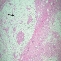
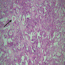
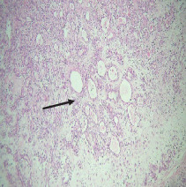
Pleomorphic adenoma (PA), the most common salivary gland tumor, presents unique challenges due to its diverse clinicopathologic features. The objective of this case series is to highlight the implication of detailed histopathological examination to guide appropriate diagnosis. This study reviews 10 cases of PA diagnosed at B.P. Koirala Institute of Health Sciences, Nepal, between 2011 and 2023. Patients ranged from 16 to 71 years, with a male-to-female ratio of 1:2.3. Most lesions (eight cases) were located on the palate, with additional cases, one on the upper lip and one on the cheek mucosa. Lesion sizes ranged from 1 to 3 cm2 and durations from 2 months to 4 years. Clinically, all lesions were well encapsulated, nontender, and nonulcerated and had normal overlying mucosa. Histopathologically, cases included classical PA, myxoid, and cellular types. Common findings were ductal structures filled with eosinophilic material, a "swarm bee" appearance, plasmacytoid cells, and myxoid stroma. Squamous differentiation and psammoma bodies were observed in some cases. No osseous or cartilaginous components were detected. All cases were excised, with no recurrences reported during at least 2 years of follow-up. Hence, proper diagnosis is crucial for effective management and long-term outcomes of PA.
期刊介绍:
Case Reports in Dentistry is a peer-reviewed, Open Access journal that publishes case reports and case series in all areas of dentistry, including periodontal diseases, dental implants, oral pathology, as well as oral and maxillofacial surgery.

 求助内容:
求助内容: 应助结果提醒方式:


