Eric C Ledbetter, Irini D Lamkin, Jeanine Peters-Kennedy
{"title":"Anterior Uveitis and Uveal Depigmentation in a Dog With Vitiligo.","authors":"Eric C Ledbetter, Irini D Lamkin, Jeanine Peters-Kennedy","doi":"10.1155/crve/6586766","DOIUrl":null,"url":null,"abstract":"<p><p><b>Objective:</b> The objective of this study is to describe the clinical and histologic features of a dog that developed anterior uveitis and uveal depigmentation in association with vitiligo. <b>Animal Studied:</b> A 3-year-old, female-spayed, Bernese Mountain Dog with a history of bilateral idiopathic anterior uveitis developed iris depigmentation, leukotrichia, and skin depigmentation. <b>Procedures:</b> The initial diagnostic evaluation for uveitis was unremarkable, including general bloodwork, urinalysis, infectious disease testing, thoracic radiographs, and abdominal ultrasound. After the development of dermatologic disease, uveodermatologic syndrome was clinically suspected and cutaneous biopsy specimens were collected for histopathology. <b>Results:</b> Cutaneous histopathology was consistent with vitiligo. Progressive and diffuse skin and hair depigmentation occurred over several years, but the dog's anterior uveitis remained well controlled on relatively minimal topical anti-inflammatory medications. No posterior segment ocular lesions developed, and the dog remained visual. <b>Conclusions and Clinical Relevance:</b> This report indicates that anterior uveitis and uveal depigmentation can develop in dogs associated with vitiligo. The presence of bilateral uveitis and uveal depigmentation, concurrent with skin and hair depigmentation, is often considered suggestive of uveodermatologic syndrome in a dog. This report illustrates the importance of cutaneous histopathology to confirm a clinical suspicion even in the most suggestive of clinical presentations.</p>","PeriodicalId":37339,"journal":{"name":"Case Reports in Veterinary Medicine","volume":"2025 ","pages":"6586766"},"PeriodicalIF":0.0000,"publicationDate":"2025-01-13","publicationTypes":"Journal Article","fieldsOfStudy":null,"isOpenAccess":false,"openAccessPdf":"https://www.ncbi.nlm.nih.gov/pmc/articles/PMC11745554/pdf/","citationCount":"0","resultStr":null,"platform":"Semanticscholar","paperid":null,"PeriodicalName":"Case Reports in Veterinary Medicine","FirstCategoryId":"1085","ListUrlMain":"https://doi.org/10.1155/crve/6586766","RegionNum":0,"RegionCategory":null,"ArticlePicture":[],"TitleCN":null,"AbstractTextCN":null,"PMCID":null,"EPubDate":"2025/1/1 0:00:00","PubModel":"eCollection","JCR":"Q3","JCRName":"Veterinary","Score":null,"Total":0}
引用次数: 0
Abstract
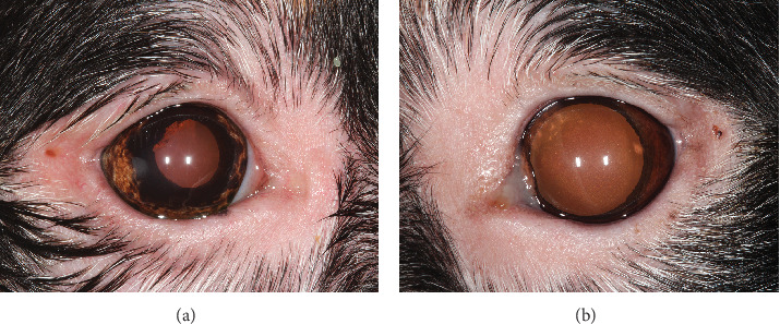
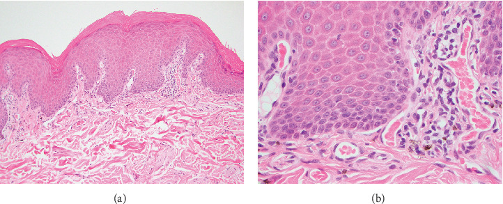
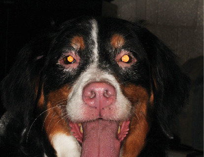
Objective: The objective of this study is to describe the clinical and histologic features of a dog that developed anterior uveitis and uveal depigmentation in association with vitiligo. Animal Studied: A 3-year-old, female-spayed, Bernese Mountain Dog with a history of bilateral idiopathic anterior uveitis developed iris depigmentation, leukotrichia, and skin depigmentation. Procedures: The initial diagnostic evaluation for uveitis was unremarkable, including general bloodwork, urinalysis, infectious disease testing, thoracic radiographs, and abdominal ultrasound. After the development of dermatologic disease, uveodermatologic syndrome was clinically suspected and cutaneous biopsy specimens were collected for histopathology. Results: Cutaneous histopathology was consistent with vitiligo. Progressive and diffuse skin and hair depigmentation occurred over several years, but the dog's anterior uveitis remained well controlled on relatively minimal topical anti-inflammatory medications. No posterior segment ocular lesions developed, and the dog remained visual. Conclusions and Clinical Relevance: This report indicates that anterior uveitis and uveal depigmentation can develop in dogs associated with vitiligo. The presence of bilateral uveitis and uveal depigmentation, concurrent with skin and hair depigmentation, is often considered suggestive of uveodermatologic syndrome in a dog. This report illustrates the importance of cutaneous histopathology to confirm a clinical suspicion even in the most suggestive of clinical presentations.
期刊介绍:
Case Reports in Veterinary Medicine is a peer-reviewed, Open Access journal that publishes case reports and case series in all areas of veterinary medicine.

 求助内容:
求助内容: 应助结果提醒方式:


