P J Stenzel, A Thomas, M Schindeldecker, S Macher-Goeppinger, S Porubsky, A Haferkamp, I Tsaur, W Roth, K E Tagscherer
{"title":"Tumor-infiltrating plasma cells are a prognostic factor in penile squamous cell carcinoma.","authors":"P J Stenzel, A Thomas, M Schindeldecker, S Macher-Goeppinger, S Porubsky, A Haferkamp, I Tsaur, W Roth, K E Tagscherer","doi":"10.1007/s00428-024-04013-1","DOIUrl":null,"url":null,"abstract":"<p><p>Penile cancer (PeCa) is a rare disease with poor prognosis in the metastatic stage. Neither effective adjuvant nor palliative therapeutic options are available. Research efforts in this field have so far failed to establish robust predictors of survival. To identify prognostic targets in PeCa, the current project focused on characterizing the tumor microenvironment (TME). A study cohort of 93 men with PeCa was used for the construction of a tissue microarray and immunohistochemical staining for CD3, CD4, CD8, CD20, CD56, CD138, FoxP3, and PD-L1. The quantity and spatial distribution of tumor-infiltrating immune cells were analyzed using digital image analysis. PD-L1 staining of tumor and immune cells was manually scored (combined positivity score (CPS)). T cells, T helper cells, cytotoxic T cells (CTLs), and regulatory T cells were detected in > 90% of PeCa and B cells in 88%, plasma cells in 85%, and NK cells in 23%. Approximately 50% of the PeCa samples were PD-L1 positive. In the univariate survival analysis, high PD-L1 CPS, plasma cells, CTLs, and B cells were significantly associated with favorable overall survival (OS), and the latter two with adverse recurrence-free survival. In multivariate analysis, plasma cells remained a significant factor for favorable OS (p = 0.04). In this study, the immune cells in the TME, especially plasma cells, were favorably associated with patient survival compared to other established prognostic factors in PeCa. Contemporarily, plasma cells have been discussed in the light of contributing to responses to modern immunotherapies. The results of this study support this notion.</p>","PeriodicalId":23514,"journal":{"name":"Virchows Archiv","volume":" ","pages":"687-699"},"PeriodicalIF":3.1000,"publicationDate":"2025-09-01","publicationTypes":"Journal Article","fieldsOfStudy":null,"isOpenAccess":false,"openAccessPdf":"https://www.ncbi.nlm.nih.gov/pmc/articles/PMC12488782/pdf/","citationCount":"0","resultStr":null,"platform":"Semanticscholar","paperid":null,"PeriodicalName":"Virchows Archiv","FirstCategoryId":"3","ListUrlMain":"https://doi.org/10.1007/s00428-024-04013-1","RegionNum":3,"RegionCategory":"医学","ArticlePicture":[],"TitleCN":null,"AbstractTextCN":null,"PMCID":null,"EPubDate":"2025/1/14 0:00:00","PubModel":"Epub","JCR":"Q1","JCRName":"PATHOLOGY","Score":null,"Total":0}
引用次数: 0
Abstract
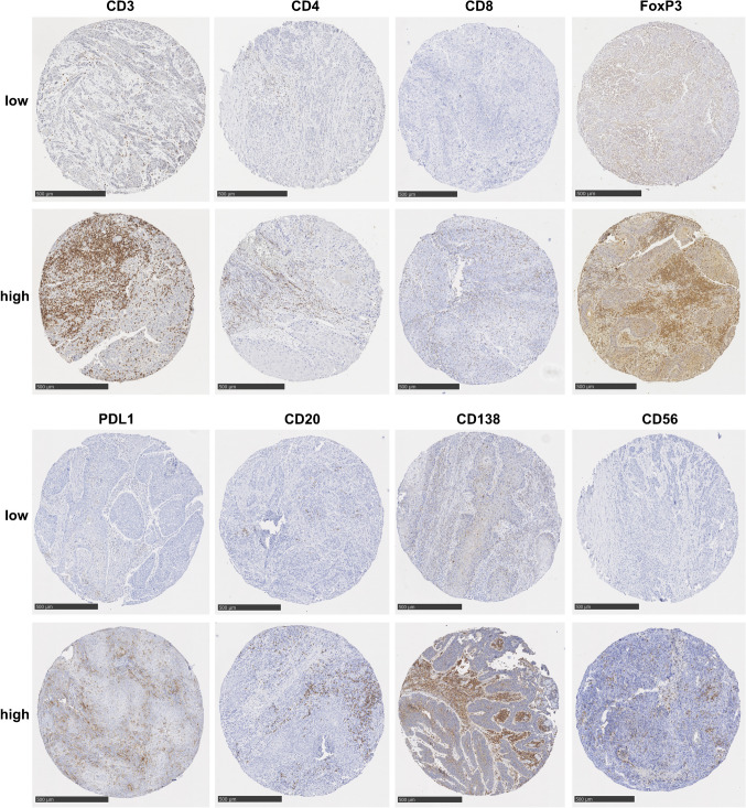
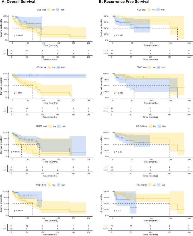
Penile cancer (PeCa) is a rare disease with poor prognosis in the metastatic stage. Neither effective adjuvant nor palliative therapeutic options are available. Research efforts in this field have so far failed to establish robust predictors of survival. To identify prognostic targets in PeCa, the current project focused on characterizing the tumor microenvironment (TME). A study cohort of 93 men with PeCa was used for the construction of a tissue microarray and immunohistochemical staining for CD3, CD4, CD8, CD20, CD56, CD138, FoxP3, and PD-L1. The quantity and spatial distribution of tumor-infiltrating immune cells were analyzed using digital image analysis. PD-L1 staining of tumor and immune cells was manually scored (combined positivity score (CPS)). T cells, T helper cells, cytotoxic T cells (CTLs), and regulatory T cells were detected in > 90% of PeCa and B cells in 88%, plasma cells in 85%, and NK cells in 23%. Approximately 50% of the PeCa samples were PD-L1 positive. In the univariate survival analysis, high PD-L1 CPS, plasma cells, CTLs, and B cells were significantly associated with favorable overall survival (OS), and the latter two with adverse recurrence-free survival. In multivariate analysis, plasma cells remained a significant factor for favorable OS (p = 0.04). In this study, the immune cells in the TME, especially plasma cells, were favorably associated with patient survival compared to other established prognostic factors in PeCa. Contemporarily, plasma cells have been discussed in the light of contributing to responses to modern immunotherapies. The results of this study support this notion.
期刊介绍:
Manuscripts of original studies reinforcing the evidence base of modern diagnostic pathology, using immunocytochemical, molecular and ultrastructural techniques, will be welcomed. In addition, papers on critical evaluation of diagnostic criteria but also broadsheets and guidelines with a solid evidence base will be considered. Consideration will also be given to reports of work in other fields relevant to the understanding of human pathology as well as manuscripts on the application of new methods and techniques in pathology. Submission of purely experimental articles is discouraged but manuscripts on experimental work applicable to diagnostic pathology are welcomed. Biomarker studies are welcomed but need to abide by strict rules (e.g. REMARK) of adequate sample size and relevant marker choice. Single marker studies on limited patient series without validated application will as a rule not be considered. Case reports will only be considered when they provide substantial new information with an impact on understanding disease or diagnostic practice.

 求助内容:
求助内容: 应助结果提醒方式:


