{"title":"The Investing Layer of the Deep Cervical Fascia: An Alternative Site for Ultrasound-guided Supraclavicular Nerve Block - A Cadaveric Anatomical Study.","authors":"Sandeep Diwan, S Shivaprakash, Rasika Timane, Pallavi Pai, Zenab Bohra, Abhijit Sukumaran Nair","doi":"10.4103/jmu.jmu_121_23","DOIUrl":null,"url":null,"abstract":"<p><strong>Background: </strong>It is very well known that the supraclavicular nerve (SCN) which occupies the inferior part of the superficial cervical plexus basically originates from the ventral rami of C2-C4, then travels caudally into the investing layer of the deep cervical fascia (IL-DCF) alternatively termed the \"prevertebral fascia.\"</p><p><strong>Methods: </strong>This cadaveric study (a total of 6 soft-embalmed cadavers and bilateral dissections, i.e. 12 specimens) intended to ascertain the location of SCN within the layers of the IL-DCF. We hypothesized that ultrasonography identification of SCN within the IL-DCF and needle tip positioned between the layers of IL-DCF provide an alternative site for the blockade of the SCN.</p><p><strong>Results: </strong>After dissection, we described a compact double-layered IL-DCF hosting the SCNs and a specific topographic arrangement at the C4 root with SCN lateral and C4 branches of the phrenic nerve medial to the C4.</p><p><strong>Conclusion: </strong>We recommend another alternative site for the SCN block at a site in the compact double layer of IL-DCF. We conclude that a caudal site at the exit of SCN from the IL-DCF would be appropriate to perform the intervention.</p>","PeriodicalId":45466,"journal":{"name":"Journal of Medical Ultrasound","volume":"32 4","pages":"318-322"},"PeriodicalIF":0.8000,"publicationDate":"2024-11-30","publicationTypes":"Journal Article","fieldsOfStudy":null,"isOpenAccess":false,"openAccessPdf":"https://www.ncbi.nlm.nih.gov/pmc/articles/PMC11717074/pdf/","citationCount":"0","resultStr":null,"platform":"Semanticscholar","paperid":null,"PeriodicalName":"Journal of Medical Ultrasound","FirstCategoryId":"1085","ListUrlMain":"https://doi.org/10.4103/jmu.jmu_121_23","RegionNum":0,"RegionCategory":null,"ArticlePicture":[],"TitleCN":null,"AbstractTextCN":null,"PMCID":null,"EPubDate":"2024/10/1 0:00:00","PubModel":"eCollection","JCR":"Q4","JCRName":"RADIOLOGY, NUCLEAR MEDICINE & MEDICAL IMAGING","Score":null,"Total":0}
引用次数: 0
Abstract
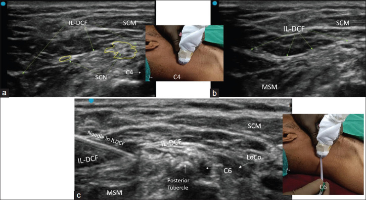
Background: It is very well known that the supraclavicular nerve (SCN) which occupies the inferior part of the superficial cervical plexus basically originates from the ventral rami of C2-C4, then travels caudally into the investing layer of the deep cervical fascia (IL-DCF) alternatively termed the "prevertebral fascia."
Methods: This cadaveric study (a total of 6 soft-embalmed cadavers and bilateral dissections, i.e. 12 specimens) intended to ascertain the location of SCN within the layers of the IL-DCF. We hypothesized that ultrasonography identification of SCN within the IL-DCF and needle tip positioned between the layers of IL-DCF provide an alternative site for the blockade of the SCN.
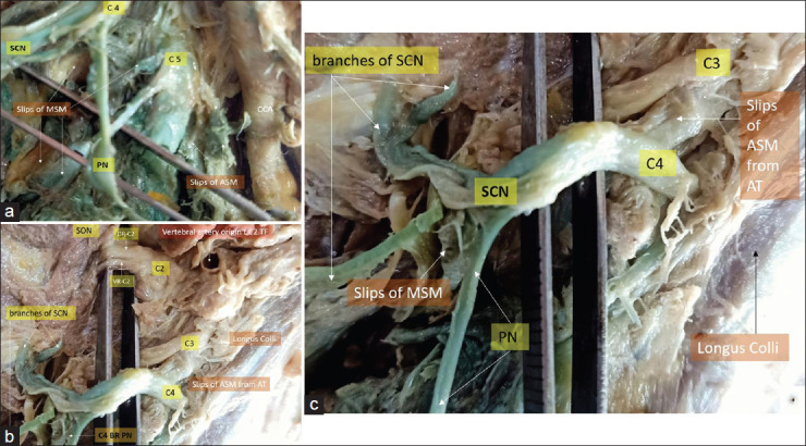
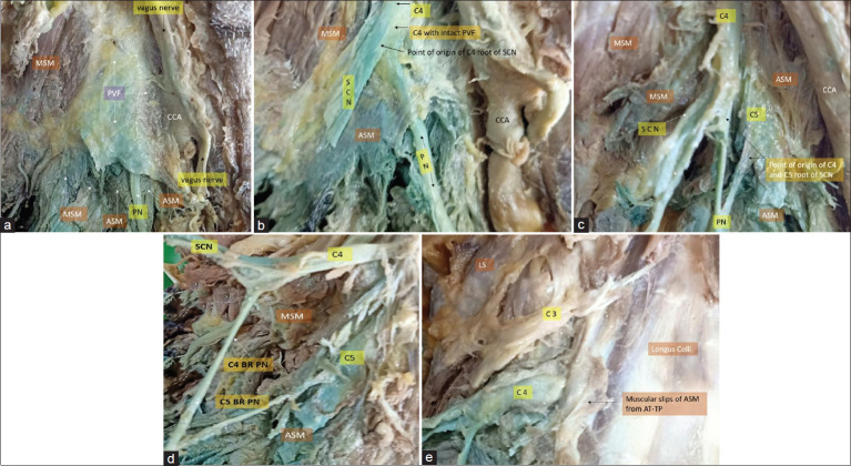
Results: After dissection, we described a compact double-layered IL-DCF hosting the SCNs and a specific topographic arrangement at the C4 root with SCN lateral and C4 branches of the phrenic nerve medial to the C4.
Conclusion: We recommend another alternative site for the SCN block at a site in the compact double layer of IL-DCF. We conclude that a caudal site at the exit of SCN from the IL-DCF would be appropriate to perform the intervention.
期刊介绍:
The Journal of Medical Ultrasound is the peer-reviewed publication of the Asian Federation of Societies for Ultrasound in Medicine and Biology, and the Chinese Taipei Society of Ultrasound in Medicine. Its aim is to promote clinical and scientific research in ultrasonography, and to serve as a channel of communication among sonologists, sonographers, and medical ultrasound physicians in the Asia-Pacific region and wider international community. The Journal invites original contributions relating to the clinical and laboratory investigations and applications of ultrasonography.

 求助内容:
求助内容: 应助结果提醒方式:


