{"title":"An atypical case of retinal pigment epithelium tear with remodeling and visual preservation.","authors":"Charles Jit Teng Ong, Chui Ming Gemmy Cheung","doi":"10.4103/tjo.TJO-D-24-00051","DOIUrl":null,"url":null,"abstract":"<p><p>This report describes a patient with polypoidal choroidal vasculopathy (PCV) with fovea-involving retinal pigment epithelium (RPE) tear that showed tissue remodeling with a good visual outcome. Imaging over the patient's clinical course from 2019 was reviewed. A 74-year-old female presented with left submacular hemorrhage and a large multi-lobular pigment epithelial detachment. Left eye vision was 6/19 at the presentation. Indocyanine green angiography (ICGA) revealed underlying PCV. One month after initiation of intravitreal aflibercept (IVA, Bayer), she developed fresh subretinal hemorrhage. An RPE tear of 1 disc area in size, centered over the fovea was diagnosed. The torn RPE edge was scrolled up temporal to the fovea on spectral-domain optical coherence tomography (SD-OCT), with hypertransmission into the choroid observed over the area of RPE loss. Left eye vision after the RPE tear was 6/15. Over the next 2 months, the subretinal hemorrhage resolved following further IVA. At month 3, fundus autofluorescence (FAF) demonstrated hypo-autofluorescence while fundus fluorescein angiography (FFA) and ICGA showed a window defect corresponding to the area of RPE tear. On SD-OCT, there was a faint hyper-reflective layer where one might expect the RPE layer to be. Serial SD-OCT scans over 5 years revealed increasing prominence of the hyperreflective layer between the ellipsoid zone and Bruch's membrane. FAF remained hypo-autofluorescent. At the last review, the patient retained 6/9 vision. We report a case of fovea-involving RPE tear documented with multimodal imaging with good visual outcome, which is atypical. Serial OCT suggests tissue remodeling may explain the functional preservation.</p>","PeriodicalId":44978,"journal":{"name":"Taiwan Journal of Ophthalmology","volume":"14 4","pages":"614-618"},"PeriodicalIF":1.2000,"publicationDate":"2024-10-29","publicationTypes":"Journal Article","fieldsOfStudy":null,"isOpenAccess":false,"openAccessPdf":"https://www.ncbi.nlm.nih.gov/pmc/articles/PMC11717326/pdf/","citationCount":"0","resultStr":null,"platform":"Semanticscholar","paperid":null,"PeriodicalName":"Taiwan Journal of Ophthalmology","FirstCategoryId":"1085","ListUrlMain":"https://doi.org/10.4103/tjo.TJO-D-24-00051","RegionNum":0,"RegionCategory":null,"ArticlePicture":[],"TitleCN":null,"AbstractTextCN":null,"PMCID":null,"EPubDate":"2024/10/1 0:00:00","PubModel":"eCollection","JCR":"Q4","JCRName":"OPHTHALMOLOGY","Score":null,"Total":0}
引用次数: 0
Abstract
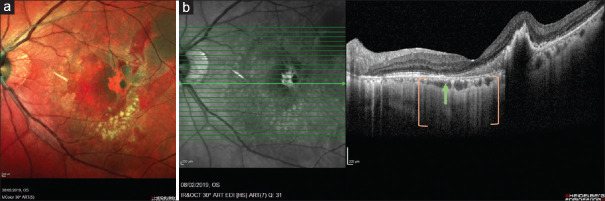
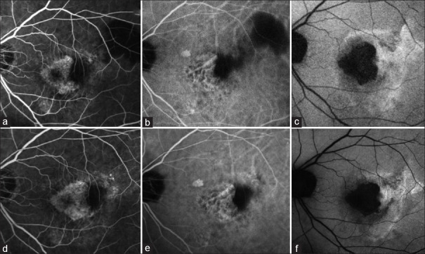
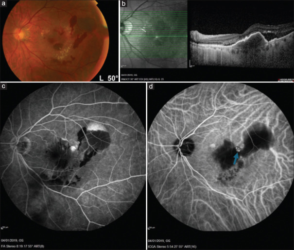
This report describes a patient with polypoidal choroidal vasculopathy (PCV) with fovea-involving retinal pigment epithelium (RPE) tear that showed tissue remodeling with a good visual outcome. Imaging over the patient's clinical course from 2019 was reviewed. A 74-year-old female presented with left submacular hemorrhage and a large multi-lobular pigment epithelial detachment. Left eye vision was 6/19 at the presentation. Indocyanine green angiography (ICGA) revealed underlying PCV. One month after initiation of intravitreal aflibercept (IVA, Bayer), she developed fresh subretinal hemorrhage. An RPE tear of 1 disc area in size, centered over the fovea was diagnosed. The torn RPE edge was scrolled up temporal to the fovea on spectral-domain optical coherence tomography (SD-OCT), with hypertransmission into the choroid observed over the area of RPE loss. Left eye vision after the RPE tear was 6/15. Over the next 2 months, the subretinal hemorrhage resolved following further IVA. At month 3, fundus autofluorescence (FAF) demonstrated hypo-autofluorescence while fundus fluorescein angiography (FFA) and ICGA showed a window defect corresponding to the area of RPE tear. On SD-OCT, there was a faint hyper-reflective layer where one might expect the RPE layer to be. Serial SD-OCT scans over 5 years revealed increasing prominence of the hyperreflective layer between the ellipsoid zone and Bruch's membrane. FAF remained hypo-autofluorescent. At the last review, the patient retained 6/9 vision. We report a case of fovea-involving RPE tear documented with multimodal imaging with good visual outcome, which is atypical. Serial OCT suggests tissue remodeling may explain the functional preservation.

 求助内容:
求助内容: 应助结果提醒方式:


