{"title":"Optical coherence tomography characteristics in hydroxychloroquine retinopathy and the correlations with visual deterioration in Taiwanese.","authors":"Shao-Kai He, Tso-Ting Lai, Yi-Ting Hsieh","doi":"10.4103/tjo.TJO-D-24-00071","DOIUrl":null,"url":null,"abstract":"<p><strong>Purpose: </strong>This study aimed to investigate optical coherence tomography (OCT) characteristics in hydroxychloroquine (HCQ) retinopathy and their correlation with visual acuity among Taiwanese patients.</p><p><strong>Materials and methods: </strong>We retrospectively recruited patients undergoing long-term HCQ treatment who had received examinations of best-corrected visual acuity and OCT scans. We observed disruptions in the ellipsoid zone (EZ) and retinal pigment epithelium (RPE) across different retinal regions. Principal component analysis (PCA) was employed to identify the most significant factors associated with visual deterioration.</p><p><strong>Results: </strong>Among the 120 eyes included in the study, HCQ retinopathy was present in 42 eyes (35.0%). In patients with mild-to-moderate retinopathy, the pericentral pattern was predominant (75.0%), whereas no parafoveal pattern was observed. Serial examinations revealed that lesions typically progressed from pericentral to parafoveal and foveal regions. EZ disruption was observed in all affected cases, most frequently at the pericentral region (100%), followed by the perifoveal (87.4%), parafoveal (72.1%), and foveal (43.2%) regions. RPE disruption was noted in 59.5% of cases, with the highest prevalence at the pericentral (53.2%) and perifoveal (52.3%) regions, followed by the parafoveal (33.3%) and foveal (28.8%) regions. PCA identified RPE disruption at the fovea and parafoveal regions as the most strongly correlated factors for visual deterioration.</p><p><strong>Conclusions: </strong>In Taiwanese patients, HCQ retinopathy predominantly manifests with pericentral lesions, while isolated parafoveal lesions are rare as an initial presentation. RPE disruption, rather than EZ disruption, appears to be the primary determinant for visual deterioration in this population.</p>","PeriodicalId":44978,"journal":{"name":"Taiwan Journal of Ophthalmology","volume":"14 4","pages":"565-572"},"PeriodicalIF":1.2000,"publicationDate":"2024-11-11","publicationTypes":"Journal Article","fieldsOfStudy":null,"isOpenAccess":false,"openAccessPdf":"https://www.ncbi.nlm.nih.gov/pmc/articles/PMC11717340/pdf/","citationCount":"0","resultStr":null,"platform":"Semanticscholar","paperid":null,"PeriodicalName":"Taiwan Journal of Ophthalmology","FirstCategoryId":"1085","ListUrlMain":"https://doi.org/10.4103/tjo.TJO-D-24-00071","RegionNum":0,"RegionCategory":null,"ArticlePicture":[],"TitleCN":null,"AbstractTextCN":null,"PMCID":null,"EPubDate":"2024/10/1 0:00:00","PubModel":"eCollection","JCR":"Q4","JCRName":"OPHTHALMOLOGY","Score":null,"Total":0}
引用次数: 0
Abstract
Purpose: This study aimed to investigate optical coherence tomography (OCT) characteristics in hydroxychloroquine (HCQ) retinopathy and their correlation with visual acuity among Taiwanese patients.
Materials and methods: We retrospectively recruited patients undergoing long-term HCQ treatment who had received examinations of best-corrected visual acuity and OCT scans. We observed disruptions in the ellipsoid zone (EZ) and retinal pigment epithelium (RPE) across different retinal regions. Principal component analysis (PCA) was employed to identify the most significant factors associated with visual deterioration.
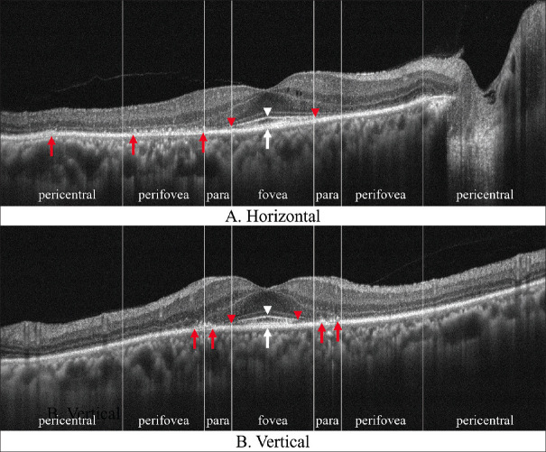
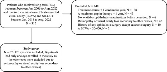
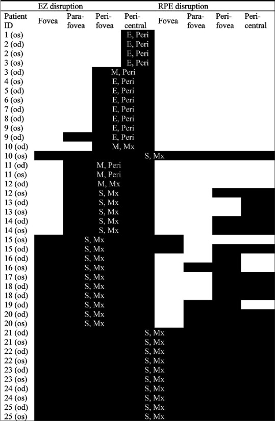
Results: Among the 120 eyes included in the study, HCQ retinopathy was present in 42 eyes (35.0%). In patients with mild-to-moderate retinopathy, the pericentral pattern was predominant (75.0%), whereas no parafoveal pattern was observed. Serial examinations revealed that lesions typically progressed from pericentral to parafoveal and foveal regions. EZ disruption was observed in all affected cases, most frequently at the pericentral region (100%), followed by the perifoveal (87.4%), parafoveal (72.1%), and foveal (43.2%) regions. RPE disruption was noted in 59.5% of cases, with the highest prevalence at the pericentral (53.2%) and perifoveal (52.3%) regions, followed by the parafoveal (33.3%) and foveal (28.8%) regions. PCA identified RPE disruption at the fovea and parafoveal regions as the most strongly correlated factors for visual deterioration.
Conclusions: In Taiwanese patients, HCQ retinopathy predominantly manifests with pericentral lesions, while isolated parafoveal lesions are rare as an initial presentation. RPE disruption, rather than EZ disruption, appears to be the primary determinant for visual deterioration in this population.

 求助内容:
求助内容: 应助结果提醒方式:


