Diane Choi, Jean-Francois Paré, Shashank Dravid, Yoland Smith
{"title":"Ultrastructural Localization of Glutamate Delta Receptor 1 in the Rodent and Primate Lateral Habenula","authors":"Diane Choi, Jean-Francois Paré, Shashank Dravid, Yoland Smith","doi":"10.1002/cne.70019","DOIUrl":null,"url":null,"abstract":"<p>Glutamate delta receptor 1 (GluD1) is a unique synaptogenic molecule expressed at excitatory and inhibitory synapses. The lateral habenula (LHb), a subcortical structure that regulates negative reward prediction error and major monoaminergic systems, is enriched in GluD1. LHb dysfunction has been implicated in psychiatric disorders such as depression and schizophrenia, both of which are associated with <i>GRID1</i>, the gene that encodes GluD1. Thus, disruption in GluD1 synaptic signaling may contribute to LHb dysfunction and the pathophysiology of LHb-associated disorders. Despite its strong cellular expression, little is known about the subsynaptic and subcellular localization of GluD1 in LHb neurons. Given that GluD1 is involved in the development and/or regulation of glutamatergic and GABAergic synapses in various brain regions, a detailed map of GluD1 synaptic localization is essential to elucidate its role in the LHb. To address this issue, we used immunoelectron microscopy methods in rodents and monkeys. In both species, GluD1 immunoreactivity was primarily expressed in dendritic profiles, with lower expression in somata, spines, and glial elements. Pre- and post-embedding immunogold experiments revealed strong GluD1 expression in the core of symmetric GABAergic synapses. Albeit less frequent, GluD1 was also found at the edges (i.e., perisynaptic) of asymmetric, putative glutamatergic synapses. Through the combination of anterograde tracing with immunogold labeling in rats, we showed that axon terminals from the entopeduncular nucleus and the lateral hypothalamus express postsynaptic GluD1 immunolabeling in the LHb. Our findings suggest that GluD1 may play a critical role in modulating GABAergic transmission in the rodent and primate LHb.</p>","PeriodicalId":15552,"journal":{"name":"Journal of Comparative Neurology","volume":"533 1","pages":""},"PeriodicalIF":2.1000,"publicationDate":"2025-01-10","publicationTypes":"Journal Article","fieldsOfStudy":null,"isOpenAccess":false,"openAccessPdf":"https://www.ncbi.nlm.nih.gov/pmc/articles/PMC11723828/pdf/","citationCount":"0","resultStr":null,"platform":"Semanticscholar","paperid":null,"PeriodicalName":"Journal of Comparative Neurology","FirstCategoryId":"3","ListUrlMain":"https://onlinelibrary.wiley.com/doi/10.1002/cne.70019","RegionNum":4,"RegionCategory":"医学","ArticlePicture":[],"TitleCN":null,"AbstractTextCN":null,"PMCID":null,"EPubDate":"","PubModel":"","JCR":"Q3","JCRName":"NEUROSCIENCES","Score":null,"Total":0}
引用次数: 0
Abstract
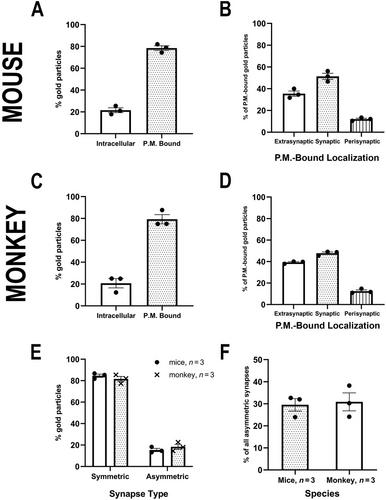
Glutamate delta receptor 1 (GluD1) is a unique synaptogenic molecule expressed at excitatory and inhibitory synapses. The lateral habenula (LHb), a subcortical structure that regulates negative reward prediction error and major monoaminergic systems, is enriched in GluD1. LHb dysfunction has been implicated in psychiatric disorders such as depression and schizophrenia, both of which are associated with GRID1, the gene that encodes GluD1. Thus, disruption in GluD1 synaptic signaling may contribute to LHb dysfunction and the pathophysiology of LHb-associated disorders. Despite its strong cellular expression, little is known about the subsynaptic and subcellular localization of GluD1 in LHb neurons. Given that GluD1 is involved in the development and/or regulation of glutamatergic and GABAergic synapses in various brain regions, a detailed map of GluD1 synaptic localization is essential to elucidate its role in the LHb. To address this issue, we used immunoelectron microscopy methods in rodents and monkeys. In both species, GluD1 immunoreactivity was primarily expressed in dendritic profiles, with lower expression in somata, spines, and glial elements. Pre- and post-embedding immunogold experiments revealed strong GluD1 expression in the core of symmetric GABAergic synapses. Albeit less frequent, GluD1 was also found at the edges (i.e., perisynaptic) of asymmetric, putative glutamatergic synapses. Through the combination of anterograde tracing with immunogold labeling in rats, we showed that axon terminals from the entopeduncular nucleus and the lateral hypothalamus express postsynaptic GluD1 immunolabeling in the LHb. Our findings suggest that GluD1 may play a critical role in modulating GABAergic transmission in the rodent and primate LHb.
期刊介绍:
Established in 1891, JCN is the oldest continually published basic neuroscience journal. Historically, as the name suggests, the journal focused on a comparison among species to uncover the intricacies of how the brain functions. In modern times, this research is called systems neuroscience where animal models are used to mimic core cognitive processes with the ultimate goal of understanding neural circuits and connections that give rise to behavioral patterns and different neural states.
Research published in JCN covers all species from invertebrates to humans, and the reports inform the readers about the function and organization of nervous systems in species with an emphasis on the way that species adaptations inform about the function or organization of the nervous systems, rather than on their evolution per se.
JCN publishes primary research articles and critical commentaries and review-type articles offering expert insight in to cutting edge research in the field of systems neuroscience; a complete list of contribution types is given in the Author Guidelines. For primary research contributions, only full-length investigative reports are desired; the journal does not accept short communications.

 求助内容:
求助内容: 应助结果提醒方式:


