{"title":"Correction to “Inhibition of ferroptosis of renal tubular cells with total flavones of Abelmoschus manihot alleviates diabetic tubulopathy”","authors":"","doi":"10.1002/ar.25617","DOIUrl":null,"url":null,"abstract":"<p>Wang, M.-Z., Cai, Y.-F., Fang, Q.-J., Liu, Y.-L., Wang, J., Chen, J.-X., Fu, Y., Wan, B.-Y., Tu, Y., Wu, W., Wan, Y.-G., & Mu, G.-L. (2023). Inhibition of ferroptosis of renal tubular cells with total flavones of <i>Abelmoschus manihot</i> alleviates diabetic tubulopathy. The Anatomical Record, 306(12), 3199–3213. https://doi.org/10.1002/ar.25123</p><p>In the originally-published article, author Yi-Gang Wan's affiliation is incorrect. The correct affiliation is:</p><p>Yi-Gang Wan<sup>1,3</sup></p><p><sup>1</sup>Department of Traditional Chinese Medicine, Nanjing Drum Tower Hospital Clinical College of Nanjing University of Chinese Medicine, Nanjing, China.</p><p><sup>3</sup>Department of Traditional Chinese Medicine, Nanjing Drum Tower Hospital, The Affiliated Hospital of Nanjing University Medical School, Nanjing, China.</p><p>Figure 7 is incorrect due to image overlap and the representative characteristics of the image being unclear. The correct Figure 7 is included below.</p><p><b>FIGURE 7</b> The ferroptosis-related changes triggered by AGEs in vitro. (a) The cell viability of the NRK-52E cells cultured in the media with various AGEs concentrations for 24 and 48 h, respectively; (b) The cell viability of the NRK-52E cells cultured in the media with various RSL-3 concentrations for 24 h; (c) FerroOrange staining in the NRK-52E cells (×800). Scale bar = 20 μm; (d) The production of ROS in the NRK-52E cells (×800). Scale bar = 20 μm; (e) The quantification of Fe<sup>2+</sup> formation fluorescence intensity; (f) The quantification of ROS formation fluorescence intensity. Data are expressed as mean ± SD. **<i>p</i> < 0.01.</p><p>Figure 8(f) is incorrect due to an overlapped image. The correct Figure 8 is included below.</p><p><b>FIGURE 8</b> The effects of TFA and dapagliflozin (Dapa) on ferroptosis-related changes in vitro. (a) The cell viability in the cultured NRK-52E cells exposed to AGEs at 200 μg/mL for 48 h with or without TFA at 5, 10, 20, and 30 μg/mL for 24 h; (b) The cell viability in the cultured NRK-52E cells exposed to AGEs at 200 μg/mL for 48 h with or without Dapa at 10, 20, 50, and 100 μM for 24 h; (c) The cell viability of the NRK-52E cells cultured in the media with various Fer-1 concentrations for 24 h; (d) The cell viability in the cultured NRK-52E cells exposed to AGEs at 200 μg/mL for 48 h with or without Fer-1 at 0.5, 1.0, 1.5, and 2.0 μM for 24 h; (e) FerroOrange staining in the NRK-52E cells (×800). Scale bar = 20 μm; (f) The production of ROS in the NRK-52E cells (×800). Scale bar = 20 μm. Data are expressed as mean ± SD. **<i>p</i> < 0.01.</p><p>We apologize for these errors.</p>","PeriodicalId":50965,"journal":{"name":"Anatomical Record-Advances in Integrative Anatomy and Evolutionary Biology","volume":"308 8","pages":"2275-2277"},"PeriodicalIF":2.1000,"publicationDate":"2025-01-09","publicationTypes":"Journal Article","fieldsOfStudy":null,"isOpenAccess":false,"openAccessPdf":"https://onlinelibrary.wiley.com/doi/epdf/10.1002/ar.25617","citationCount":"0","resultStr":null,"platform":"Semanticscholar","paperid":null,"PeriodicalName":"Anatomical Record-Advances in Integrative Anatomy and Evolutionary Biology","FirstCategoryId":"3","ListUrlMain":"https://anatomypubs.onlinelibrary.wiley.com/doi/10.1002/ar.25617","RegionNum":4,"RegionCategory":"医学","ArticlePicture":[],"TitleCN":null,"AbstractTextCN":null,"PMCID":null,"EPubDate":"","PubModel":"","JCR":"Q2","JCRName":"ANATOMY & MORPHOLOGY","Score":null,"Total":0}
引用次数: 0
Abstract
Wang, M.-Z., Cai, Y.-F., Fang, Q.-J., Liu, Y.-L., Wang, J., Chen, J.-X., Fu, Y., Wan, B.-Y., Tu, Y., Wu, W., Wan, Y.-G., & Mu, G.-L. (2023). Inhibition of ferroptosis of renal tubular cells with total flavones of Abelmoschus manihot alleviates diabetic tubulopathy. The Anatomical Record, 306(12), 3199–3213. https://doi.org/10.1002/ar.25123
In the originally-published article, author Yi-Gang Wan's affiliation is incorrect. The correct affiliation is:
Yi-Gang Wan1,3
1Department of Traditional Chinese Medicine, Nanjing Drum Tower Hospital Clinical College of Nanjing University of Chinese Medicine, Nanjing, China.
3Department of Traditional Chinese Medicine, Nanjing Drum Tower Hospital, The Affiliated Hospital of Nanjing University Medical School, Nanjing, China.
Figure 7 is incorrect due to image overlap and the representative characteristics of the image being unclear. The correct Figure 7 is included below.
FIGURE 7 The ferroptosis-related changes triggered by AGEs in vitro. (a) The cell viability of the NRK-52E cells cultured in the media with various AGEs concentrations for 24 and 48 h, respectively; (b) The cell viability of the NRK-52E cells cultured in the media with various RSL-3 concentrations for 24 h; (c) FerroOrange staining in the NRK-52E cells (×800). Scale bar = 20 μm; (d) The production of ROS in the NRK-52E cells (×800). Scale bar = 20 μm; (e) The quantification of Fe2+ formation fluorescence intensity; (f) The quantification of ROS formation fluorescence intensity. Data are expressed as mean ± SD. **p < 0.01.
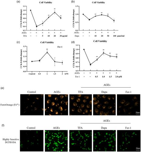
Figure 8(f) is incorrect due to an overlapped image. The correct Figure 8 is included below.
FIGURE 8 The effects of TFA and dapagliflozin (Dapa) on ferroptosis-related changes in vitro. (a) The cell viability in the cultured NRK-52E cells exposed to AGEs at 200 μg/mL for 48 h with or without TFA at 5, 10, 20, and 30 μg/mL for 24 h; (b) The cell viability in the cultured NRK-52E cells exposed to AGEs at 200 μg/mL for 48 h with or without Dapa at 10, 20, 50, and 100 μM for 24 h; (c) The cell viability of the NRK-52E cells cultured in the media with various Fer-1 concentrations for 24 h; (d) The cell viability in the cultured NRK-52E cells exposed to AGEs at 200 μg/mL for 48 h with or without Fer-1 at 0.5, 1.0, 1.5, and 2.0 μM for 24 h; (e) FerroOrange staining in the NRK-52E cells (×800). Scale bar = 20 μm; (f) The production of ROS in the NRK-52E cells (×800). Scale bar = 20 μm. Data are expressed as mean ± SD. **p < 0.01.


 求助内容:
求助内容: 应助结果提醒方式:


