Muhammad Usman, Hafiz Muhammad Rizwan, Muhammad Sohail Sajid, Razia Kausar, Urfa Bin Tahir, Haider Abbas, Muhammad Khalil Ateeq, Mohsin Raza, Mahvish Maqbool, Dalia Fouad, Farid S Ataya
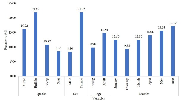
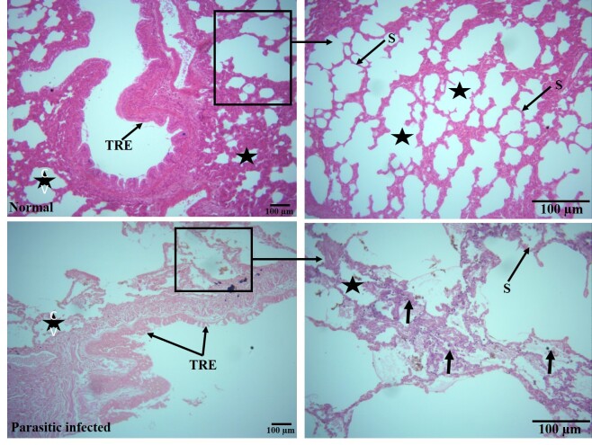
{"title":"Prevalence and histopathological analysis of cystic echinococcosis in ruminants of District Narowal, Pakistan: focus on pulmonary involvement.","authors":"Muhammad Usman, Hafiz Muhammad Rizwan, Muhammad Sohail Sajid, Razia Kausar, Urfa Bin Tahir, Haider Abbas, Muhammad Khalil Ateeq, Mohsin Raza, Mahvish Maqbool, Dalia Fouad, Farid S Ataya","doi":"10.1590/S1984-29612024080","DOIUrl":null,"url":null,"abstract":"<p><p>A total of 384 animals (sheep, goat, cattle, and buffalo) were examined for the presence of hydatid cysts only in the lungs. The lung tissue samples associated with the hydatid cyst were collected immediately after slaughter, followed by fixation in 10% formalin. The fixed tissue was subjected to paraffin embedding technique. Tissue sections of 5 microns were cut by microtome and stained using Harri's Haematoxilin and Eosin method. Overall, 13.80% of ruminants were found positive for lung infections with hydatid cyst. Only the sex of ruminants showed significant (P < 0.05) association with the infection of hydatid cyst in lungs. All other variables, such as species of ruminants, age, and months showed non-significant (P > 0.05) association. Pulmonary sections taken from infected animals revealed laminated membranes encased in a region with significant (P < 0.05) cellular infiltration (53.4 ± 7.9 µm2), primarily composed of lymphocytes, plasma cells, macrophages, and occasionally neutrophils, and eosinophils. In addition, significant (P < 0.05) epithelial disruption in the bronchioles (0.94 ± 0.05 µm2) and alveolar septa were also noticed in sections. These histopathological findings lead to the conclusion that pathological changes occur in the tissues surrounding the cyst as well as in areas more distant from the cyst.</p>","PeriodicalId":48990,"journal":{"name":"Revista Brasileira De Parasitologia Veterinaria","volume":"33 4","pages":"e016824"},"PeriodicalIF":1.2000,"publicationDate":"2024-12-20","publicationTypes":"Journal Article","fieldsOfStudy":null,"isOpenAccess":false,"openAccessPdf":"https://www.ncbi.nlm.nih.gov/pmc/articles/PMC11758849/pdf/","citationCount":"0","resultStr":null,"platform":"Semanticscholar","paperid":null,"PeriodicalName":"Revista Brasileira De Parasitologia Veterinaria","FirstCategoryId":"97","ListUrlMain":"https://doi.org/10.1590/S1984-29612024080","RegionNum":4,"RegionCategory":"农林科学","ArticlePicture":[],"TitleCN":null,"AbstractTextCN":null,"PMCID":null,"EPubDate":"2024/1/1 0:00:00","PubModel":"eCollection","JCR":"Q2","JCRName":"Veterinary","Score":null,"Total":0}
引用次数: 0
Abstract
A total of 384 animals (sheep, goat, cattle, and buffalo) were examined for the presence of hydatid cysts only in the lungs. The lung tissue samples associated with the hydatid cyst were collected immediately after slaughter, followed by fixation in 10% formalin. The fixed tissue was subjected to paraffin embedding technique. Tissue sections of 5 microns were cut by microtome and stained using Harri's Haematoxilin and Eosin method. Overall, 13.80% of ruminants were found positive for lung infections with hydatid cyst. Only the sex of ruminants showed significant (P < 0.05) association with the infection of hydatid cyst in lungs. All other variables, such as species of ruminants, age, and months showed non-significant (P > 0.05) association. Pulmonary sections taken from infected animals revealed laminated membranes encased in a region with significant (P < 0.05) cellular infiltration (53.4 ± 7.9 µm2), primarily composed of lymphocytes, plasma cells, macrophages, and occasionally neutrophils, and eosinophils. In addition, significant (P < 0.05) epithelial disruption in the bronchioles (0.94 ± 0.05 µm2) and alveolar septa were also noticed in sections. These histopathological findings lead to the conclusion that pathological changes occur in the tissues surrounding the cyst as well as in areas more distant from the cyst.
期刊介绍:
La revista es un órgano de difusión del Colegio Brasileño de Parasitología Veterinaria, con una especificidad dentro de esa área, la difusión de los resultados de la investigación brasileña en las áreas de Helmintología, Protozoología, Entomología y agentes transmitidos por artrópodos, relacionados con la salud animal.
 求助内容:
求助内容: 应助结果提醒方式:


