Yuran Huang, Zitong Ye, Zhimin Zhang, Hanchu Ye, Liang Xu, Xiang Hao, Yubing Han, Cuifang Kuang, Xu Liu
{"title":"Spatial Phasor Analysis for Optical Sectioning Nanoscopy","authors":"Yuran Huang, Zitong Ye, Zhimin Zhang, Hanchu Ye, Liang Xu, Xiang Hao, Yubing Han, Cuifang Kuang, Xu Liu","doi":"10.1021/acsphotonics.4c02336","DOIUrl":null,"url":null,"abstract":"Super-resolution microscopy has broken the traditional resolution barrier of optical microscopy. However, its application in imaging live and thick specimens has been limited. To date, optical sectioning in super-resolution microscopy either rely on inaccurate background estimation or been hindered in live-cell imaging by excessive complexity and cost. Here, we report spatial phasor image scanning microscopy (spISM), which aims to enhance the optical sectioning by a factor of ∼2 without drawbacks for any microscope equipped with a detector array. By incorporating spatial-domain phasor analysis into image scanning microscopy, spISM decodes information about the axial position, thus accurately identifying in-focus and out-of-focus signals. We demonstrate that this approach is automatic, adaptive, and robust to specimen and microscope setups. It has a rapid processing speed, enabling multicolor imaging in live-cell. The performance of the reported approach is validated by imaging up to eight subcellular structures. As spISM is fully compatible with laser scanning microscopy, it holds great potential to become a turn-key solution for biological research.","PeriodicalId":23,"journal":{"name":"ACS Photonics","volume":"130 1","pages":""},"PeriodicalIF":6.5000,"publicationDate":"2025-01-10","publicationTypes":"Journal Article","fieldsOfStudy":null,"isOpenAccess":false,"openAccessPdf":"","citationCount":"0","resultStr":null,"platform":"Semanticscholar","paperid":null,"PeriodicalName":"ACS Photonics","FirstCategoryId":"101","ListUrlMain":"https://doi.org/10.1021/acsphotonics.4c02336","RegionNum":1,"RegionCategory":"物理与天体物理","ArticlePicture":[],"TitleCN":null,"AbstractTextCN":null,"PMCID":null,"EPubDate":"","PubModel":"","JCR":"Q1","JCRName":"MATERIALS SCIENCE, MULTIDISCIPLINARY","Score":null,"Total":0}
引用次数: 0
Abstract
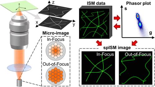
Super-resolution microscopy has broken the traditional resolution barrier of optical microscopy. However, its application in imaging live and thick specimens has been limited. To date, optical sectioning in super-resolution microscopy either rely on inaccurate background estimation or been hindered in live-cell imaging by excessive complexity and cost. Here, we report spatial phasor image scanning microscopy (spISM), which aims to enhance the optical sectioning by a factor of ∼2 without drawbacks for any microscope equipped with a detector array. By incorporating spatial-domain phasor analysis into image scanning microscopy, spISM decodes information about the axial position, thus accurately identifying in-focus and out-of-focus signals. We demonstrate that this approach is automatic, adaptive, and robust to specimen and microscope setups. It has a rapid processing speed, enabling multicolor imaging in live-cell. The performance of the reported approach is validated by imaging up to eight subcellular structures. As spISM is fully compatible with laser scanning microscopy, it holds great potential to become a turn-key solution for biological research.
期刊介绍:
Published as soon as accepted and summarized in monthly issues, ACS Photonics will publish Research Articles, Letters, Perspectives, and Reviews, to encompass the full scope of published research in this field.