Gallbladder-derived retinoic acid signalling drives reconstruction of the damaged intrahepatic biliary ducts
IF 17.3
1区 生物学
Q1 CELL BIOLOGY
引用次数: 0
Abstract
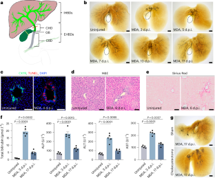
Severe damage to the intrahepatic biliary duct (IHBD) network occurs in multiple human advanced cholangiopathies, such as primary sclerosing cholangitis, biliary atresia and end-stage primary biliary cholangitis. Whether and how a severely damaged IHBD network could reconstruct has remained unclear. Here we show that, although the gallbladder is not directly connected to the IHBD, there is a common hepatic duct (CHD) in between, and severe damage to the IHBD network induces migration of gallbladder smooth muscle cells (SMCs) to coat the CHD in mouse and zebrafish models. These gallbladder-derived, CHD-coating SMCs produce retinoic acid to activate Sox9b in the CHD, which drives proliferation and ingrowth of CHD cells into the inner liver to reconstruct the IHBD network. This study reveals a hitherto unappreciated function of the gallbladder in the recovery of injured liver, and characterizes mechanisms involved in how the gallbladder and liver communicate through inter-organ cell migration to drive tissue regeneration. Carrying out cholecystectomy will thus cause previously unexpected impairments to liver health. Luo et al. show in zebrafish and mouse that, upon intrahepatic biliary duct damage, gallbladder smooth muscle cells migrate to the common hepatic duct, where they produce retinoic acid to promote regeneration of the intrahepatic biliary duct.

胆囊源性维甲酸信号驱动受损肝内胆管的重建
肝内胆管(IHBD)网络的严重损害发生在多种人类晚期胆管疾病中,如原发性硬化性胆管炎、胆道闭锁和终末期原发性胆管炎。严重受损的IHBD网络能否以及如何重建仍不清楚。本研究表明,尽管胆囊与IHBD没有直接连接,但两者之间存在肝总管(CHD),在小鼠和斑马鱼模型中,IHBD网络的严重损伤会诱导胆囊平滑肌细胞(SMCs)迁移至CHD。这些胆囊来源的、包覆冠心病的SMCs产生维甲酸,激活冠心病中的Sox9b,从而驱动冠心病细胞的增殖和长入肝脏内部,重建IHBD网络。本研究揭示了迄今为止未被认识到的胆囊在损伤肝脏恢复中的功能,并描述了胆囊和肝脏如何通过器官间细胞迁移来驱动组织再生的机制。因此,进行胆囊切除术将对肝脏健康造成先前意想不到的损害。
本文章由计算机程序翻译,如有差异,请以英文原文为准。
求助全文
约1分钟内获得全文
求助全文
来源期刊
Nature Cell Biology
生物-细胞生物学
CiteScore
28.40
自引率
0.90%
发文量
219
审稿时长
3 months
期刊介绍:
Nature Cell Biology, a prestigious journal, upholds a commitment to publishing papers of the highest quality across all areas of cell biology, with a particular focus on elucidating mechanisms underlying fundamental cell biological processes. The journal's broad scope encompasses various areas of interest, including but not limited to:
-Autophagy
-Cancer biology
-Cell adhesion and migration
-Cell cycle and growth
-Cell death
-Chromatin and epigenetics
-Cytoskeletal dynamics
-Developmental biology
-DNA replication and repair
-Mechanisms of human disease
-Mechanobiology
-Membrane traffic and dynamics
-Metabolism
-Nuclear organization and dynamics
-Organelle biology
-Proteolysis and quality control
-RNA biology
-Signal transduction
-Stem cell biology
 求助内容:
求助内容: 应助结果提醒方式:


