Clinicopathological comparison and cytokeratin-10 expression between Lichen planus and oral lichenoid lesions

IF 2.2 4区 医学 Q2 DENTISTRY, ORAL SURGERY & MEDICINE
Milena Duarte Moreira, Luanna Canal Pereira, Tahyna Duda Deps, Tânia Regina Grão Velloso, Letícia Nogueira da Gama-de-Souza, Liliana Aparecida Pimenta-Barros, Danielle Resende Camisasca
{"title":"Clinicopathological comparison and cytokeratin-10 expression between Lichen planus and oral lichenoid lesions","authors":"Milena Duarte Moreira,&nbsp;Luanna Canal Pereira,&nbsp;Tahyna Duda Deps,&nbsp;Tânia Regina Grão Velloso,&nbsp;Letícia Nogueira da Gama-de-Souza,&nbsp;Liliana Aparecida Pimenta-Barros,&nbsp;Danielle Resende Camisasca","doi":"10.1016/j.archoralbio.2024.106166","DOIUrl":null,"url":null,"abstract":"<div><h3>Objectives</h3><div>This study aimed to compare clinicopathological features and immunostaining for cytokeratin-10 between oral lichen planus and oral lichenoid lesions.</div></div><div><h3>Design</h3><div>This was a retrospective longitudinal study comparing lichen planus and oral lichenoid lesions diagnosed at the Oral Pathological Anatomy Service that analyzed sociodemographic and clinicopathological data and CK10 expression. Chi-square tests, Fisher's exact tests and Mann-Whitney tests or Student's t tests were used when appropriate, and p values &lt; 0.05 were considered significant.</div></div><div><h3>Results</h3><div>A total of 23 lichen planus and 23 lichenoid lesions were included. There was an association between oral lichen planus and symptomatology (p = 0.031). The buccal mucosa was the most affected site in both groups: 20 patients (87.0 %) showed oral lichen planus, and 16 patients (69.6 %) oral lichenoid lesions. Bilateral (p &lt; 0.001) striae (p = 0.004) are more characteristic of oral lichen planus. Oral lichen planus was associated with degeneration of the basal layer (p = 0.049) and with mild epithelial dysplasia (p &lt; 0.001). Cytokeratin-10 immunostaining was similar between the groups.</div></div><div><h3>Conclusions</h3><div>A continuous follow-up is necessary to identify different patterns of malignant transformation between groups of lesions, as well as for comparisons with lesions with a higher malignant transformation rate.</div></div>","PeriodicalId":8288,"journal":{"name":"Archives of oral biology","volume":"171 ","pages":"Article 106166"},"PeriodicalIF":2.2000,"publicationDate":"2024-12-25","publicationTypes":"Journal Article","fieldsOfStudy":null,"isOpenAccess":false,"openAccessPdf":"","citationCount":"0","resultStr":null,"platform":"Semanticscholar","paperid":null,"PeriodicalName":"Archives of oral biology","FirstCategoryId":"3","ListUrlMain":"https://www.sciencedirect.com/science/article/pii/S0003996924002875","RegionNum":4,"RegionCategory":"医学","ArticlePicture":[],"TitleCN":null,"AbstractTextCN":null,"PMCID":null,"EPubDate":"","PubModel":"","JCR":"Q2","JCRName":"DENTISTRY, ORAL SURGERY & MEDICINE","Score":null,"Total":0}
引用次数: 0

Abstract

Objectives

This study aimed to compare clinicopathological features and immunostaining for cytokeratin-10 between oral lichen planus and oral lichenoid lesions.

Design

This was a retrospective longitudinal study comparing lichen planus and oral lichenoid lesions diagnosed at the Oral Pathological Anatomy Service that analyzed sociodemographic and clinicopathological data and CK10 expression. Chi-square tests, Fisher's exact tests and Mann-Whitney tests or Student's t tests were used when appropriate, and p values < 0.05 were considered significant.

Results

A total of 23 lichen planus and 23 lichenoid lesions were included. There was an association between oral lichen planus and symptomatology (p = 0.031). The buccal mucosa was the most affected site in both groups: 20 patients (87.0 %) showed oral lichen planus, and 16 patients (69.6 %) oral lichenoid lesions. Bilateral (p < 0.001) striae (p = 0.004) are more characteristic of oral lichen planus. Oral lichen planus was associated with degeneration of the basal layer (p = 0.049) and with mild epithelial dysplasia (p < 0.001). Cytokeratin-10 immunostaining was similar between the groups.

Conclusions

A continuous follow-up is necessary to identify different patterns of malignant transformation between groups of lesions, as well as for comparisons with lesions with a higher malignant transformation rate.
扁平苔藓与口腔苔藓样病变的临床病理比较及细胞角蛋白-10的表达。
目的:比较口腔扁平苔藓和口腔类苔藓病变的临床病理特征和细胞角蛋白-10免疫染色。设计:这是一项回顾性的纵向研究,比较了口腔病理解剖服务诊断的扁平苔藓和口腔苔藓样病变,分析了社会人口统计学和临床病理数据以及CK10的表达。适当时采用卡方检验、Fisher精确检验和Mann-Whitney检验或Student’st检验,p值结果:共纳入23例扁平苔藓和23例地衣样病变。口腔扁平苔藓与症状有相关性(p = 0.031)。两组患者均以口腔黏膜为最主要受损伤部位:口腔扁平苔藓20例(87.0 %),口腔类地衣病变16例(69.6% %)。结论:持续的随访是必要的,以确定不同组间病变的不同恶性转化模式,并与恶性转化率较高的病变进行比较。
本文章由计算机程序翻译,如有差异,请以英文原文为准。
求助全文
约1分钟内获得全文 求助全文
来源期刊
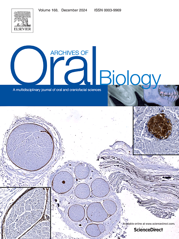
Archives of oral biology
Archives of oral biology 医学-牙科与口腔外科
CiteScore
5.10
自引率
3.30%
发文量
177
审稿时长
26 days
期刊介绍: Archives of Oral Biology is an international journal which aims to publish papers of the highest scientific quality in the oral and craniofacial sciences. The journal is particularly interested in research which advances knowledge in the mechanisms of craniofacial development and disease, including: Cell and molecular biology Molecular genetics Immunology Pathogenesis Cellular microbiology Embryology Syndromology Forensic dentistry
×
引用
GB/T 7714-2015
复制
MLA
复制
APA
复制
导出至
BibTeX EndNote RefMan NoteFirst NoteExpress
×
提示
您的信息不完整,为了账户安全,请先补充。
现在去补充
×
提示
您因"违规操作"
具体请查看互助需知
我知道了
×
提示
确定
请完成安全验证×
copy
已复制链接
快去分享给好友吧!
我知道了
右上角分享
点击右上角分享
0
联系我们:info@booksci.cn Book学术提供免费学术资源搜索服务,方便国内外学者检索中英文文献。致力于提供最便捷和优质的服务体验。 Copyright © 2023 布克学术 All rights reserved.
京ICP备2023020795号-1
ghs 京公网安备 11010802042870号
Book学术文献互助
Book学术文献互助群
群 号:481959085
Book学术官方微信