Comparison of diagnostic performance of [68Ga]-Ga-FAPI-46 and [18F]-FDG PET/CT imaging for the detection of lesions and disease staging in patients with breast cancer.
{"title":"Comparison of diagnostic performance of [<sup>68</sup>Ga]-Ga-FAPI-46 and [<sup>18</sup>F]-FDG PET/CT imaging for the detection of lesions and disease staging in patients with breast cancer.","authors":"Kiana Radmehr, Saeed Farzanefar, Mehrshad Abbasi, Yalda Salehi, Najme Karamzade-Ziarati, Alireza Emami-Ardekani, Reyhaneh Manafi-Farid, Nasim Vahidfar, Davood Beiki","doi":"10.22038/aojnmb.2024.80845.1573","DOIUrl":null,"url":null,"abstract":"<p><strong>Objectives: </strong>To compare the diagnostic performance of [<sup>68</sup>Ga]-Ga-FAPI-46 and [<sup>18</sup>F]-FDG PET/CT imaging for the detection of lesions and disease staging in breast cancer.</p><p><strong>Methods: </strong>Twelve female patients with breast cancer (mean age= 49.2±13.29 years) and previous [<sup>18</sup>F]-FDG PET/CT were recruited in the study. [<sup>68</sup>Ga]Ga-FAPI-46 imaging performed in all patients within one month after [<sup>18</sup>F]-FDG PET/CT imaging. The acquired PET/CT data with both tracers were reconstructed. Tracer avid lesions with each PET tracer were identified and the semi-quantitative parameters i.e. SUV<sub>max</sub>, lesion counts and target-to-background ratio (TBR<sub>max</sub>) were analyzed.</p><p><strong>Results: </strong>Physiologic distribution of [<sup>68</sup>Ga]-Ga-FAPI-46 was observed in the liver, blood pool and kidneys, whereas no tracer uptake was noted in the brain and heart. The mean liver SUV<sub>max</sub> for [<sup>68</sup>Ga] Ga-FAPI-46 was 1.5±0.1 which was lower than that noted for [<sup>18</sup>F]-FDG PET/CT (2.9±0.2). Likewise, the mean blood pool SUV<sub>max</sub> value for [<sup>68</sup>Ga]-Ga-FAPI-46 was lower than [<sup>18</sup>F]-FDG PET/CT (1.7±0.1 versus 2.0±0.1). [<sup>68</sup>Ga]-Ga-FAPI-46 PET/CT demonstrated higher tracer uptake in the lesions detected in the brain, bone, internal mammary and lymph nodes in 4/12 patients. The overall lesions detections and the mean SUV<sub>max</sub> values did not differ significantly between the two techniques. On the other hand, [<sup>68</sup>Ga]-Ga-FAPI-46 demonstrated higher mean TBR<sub>max</sub> than [<sup>18</sup>F] FDG PET/CT particularly for lesions detected in kidneys, chest wall, mediastinum, and musculoskeletal lesions. However, both techniques offered identical TNM staging.</p><p><strong>Conclusion: </strong>The findings of this preliminary study demonstrated that [<sup>68</sup>Ga]-Ga-FAPI-46 and [<sup>18</sup>F]-FDG PET/CT offered identical disease staging in the breast cancer patients. [<sup>68</sup>Ga]-Ga-FAPI-46 showed lower liver and blood pool uptake and an enhanced tumor-to-background ratio, thereby suggesting its potential for improved lesions detection. This may open opportunity for emerging FAP based radioligand for therapeutic applications in advanced stage breast cancers. However, this needs validation in a larger number of patients.</p>","PeriodicalId":8503,"journal":{"name":"Asia Oceania Journal of Nuclear Medicine and Biology","volume":"13 1","pages":"1-9"},"PeriodicalIF":0.0000,"publicationDate":"2025-01-01","publicationTypes":"Journal Article","fieldsOfStudy":null,"isOpenAccess":false,"openAccessPdf":"https://www.ncbi.nlm.nih.gov/pmc/articles/PMC11682476/pdf/","citationCount":"0","resultStr":null,"platform":"Semanticscholar","paperid":null,"PeriodicalName":"Asia Oceania Journal of Nuclear Medicine and Biology","FirstCategoryId":"1085","ListUrlMain":"https://doi.org/10.22038/aojnmb.2024.80845.1573","RegionNum":0,"RegionCategory":null,"ArticlePicture":[],"TitleCN":null,"AbstractTextCN":null,"PMCID":null,"EPubDate":"","PubModel":"","JCR":"Q3","JCRName":"Medicine","Score":null,"Total":0}
引用次数: 0
Abstract
Objectives: To compare the diagnostic performance of [68Ga]-Ga-FAPI-46 and [18F]-FDG PET/CT imaging for the detection of lesions and disease staging in breast cancer.
Methods: Twelve female patients with breast cancer (mean age= 49.2±13.29 years) and previous [18F]-FDG PET/CT were recruited in the study. [68Ga]Ga-FAPI-46 imaging performed in all patients within one month after [18F]-FDG PET/CT imaging. The acquired PET/CT data with both tracers were reconstructed. Tracer avid lesions with each PET tracer were identified and the semi-quantitative parameters i.e. SUVmax, lesion counts and target-to-background ratio (TBRmax) were analyzed.
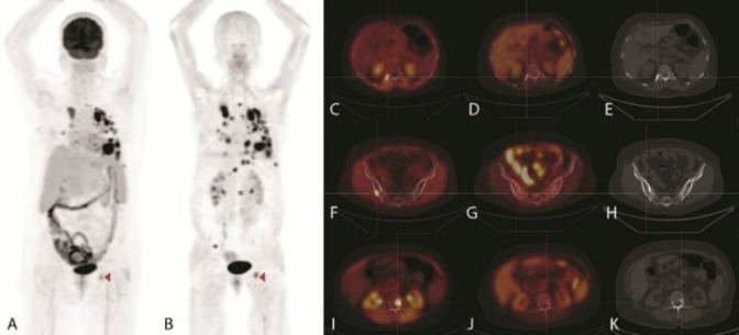
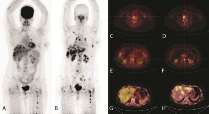
Results: Physiologic distribution of [68Ga]-Ga-FAPI-46 was observed in the liver, blood pool and kidneys, whereas no tracer uptake was noted in the brain and heart. The mean liver SUVmax for [68Ga] Ga-FAPI-46 was 1.5±0.1 which was lower than that noted for [18F]-FDG PET/CT (2.9±0.2). Likewise, the mean blood pool SUVmax value for [68Ga]-Ga-FAPI-46 was lower than [18F]-FDG PET/CT (1.7±0.1 versus 2.0±0.1). [68Ga]-Ga-FAPI-46 PET/CT demonstrated higher tracer uptake in the lesions detected in the brain, bone, internal mammary and lymph nodes in 4/12 patients. The overall lesions detections and the mean SUVmax values did not differ significantly between the two techniques. On the other hand, [68Ga]-Ga-FAPI-46 demonstrated higher mean TBRmax than [18F] FDG PET/CT particularly for lesions detected in kidneys, chest wall, mediastinum, and musculoskeletal lesions. However, both techniques offered identical TNM staging.
Conclusion: The findings of this preliminary study demonstrated that [68Ga]-Ga-FAPI-46 and [18F]-FDG PET/CT offered identical disease staging in the breast cancer patients. [68Ga]-Ga-FAPI-46 showed lower liver and blood pool uptake and an enhanced tumor-to-background ratio, thereby suggesting its potential for improved lesions detection. This may open opportunity for emerging FAP based radioligand for therapeutic applications in advanced stage breast cancers. However, this needs validation in a larger number of patients.

 求助内容:
求助内容: 应助结果提醒方式:


