Molecular basis of proton sensing by G protein-coupled receptors

IF 45.5 1区 生物学 Q1 BIOCHEMISTRY & MOLECULAR BIOLOGY
Cell Pub Date : 2025-01-02 DOI:10.1016/j.cell.2024.11.036
Matthew K. Howard, Nicholas Hoppe, Xi-Ping Huang, Darko Mitrovic, Christian B. Billesbølle, Christian B. Macdonald, Eshan Mehrotra, Patrick Rockefeller Grimes, Donovan D. Trinidad, Lucie Delemotte, Justin G. English, Willow Coyote-Maestas, Aashish Manglik
{"title":"Molecular basis of proton sensing by G protein-coupled receptors","authors":"Matthew K. Howard, Nicholas Hoppe, Xi-Ping Huang, Darko Mitrovic, Christian B. Billesbølle, Christian B. Macdonald, Eshan Mehrotra, Patrick Rockefeller Grimes, Donovan D. Trinidad, Lucie Delemotte, Justin G. English, Willow Coyote-Maestas, Aashish Manglik","doi":"10.1016/j.cell.2024.11.036","DOIUrl":null,"url":null,"abstract":"Three proton-sensing G protein-coupled receptors (GPCRs)—GPR4, GPR65, and GPR68—respond to extracellular pH to regulate diverse physiology. How protons activate these receptors is poorly understood. We determined cryogenic-electron microscopy (cryo-EM) structures of each receptor to understand the spatial arrangement of proton-sensing residues. Using deep mutational scanning (DMS), we determined the functional importance of every residue in GPR68 activation by generating ∼9,500 mutants and measuring their effects on signaling and surface expression. Constant-pH molecular dynamics simulations provided insights into the conformational landscape and protonation patterns of key residues. This unbiased approach revealed that, unlike other proton-sensitive channels and receptors, no single site is critical for proton recognition. Instead, a network of titratable residues extends from the extracellular surface to the transmembrane region, converging on canonical motifs to activate proton-sensing GPCRs. Our approach integrating structure, simulations, and unbiased functional interrogation provides a framework for understanding GPCR signaling complexity.","PeriodicalId":9656,"journal":{"name":"Cell","volume":"92 1","pages":""},"PeriodicalIF":45.5000,"publicationDate":"2025-01-02","publicationTypes":"Journal Article","fieldsOfStudy":null,"isOpenAccess":false,"openAccessPdf":"","citationCount":"0","resultStr":null,"platform":"Semanticscholar","paperid":null,"PeriodicalName":"Cell","FirstCategoryId":"99","ListUrlMain":"https://doi.org/10.1016/j.cell.2024.11.036","RegionNum":1,"RegionCategory":"生物学","ArticlePicture":[],"TitleCN":null,"AbstractTextCN":null,"PMCID":null,"EPubDate":"","PubModel":"","JCR":"Q1","JCRName":"BIOCHEMISTRY & MOLECULAR BIOLOGY","Score":null,"Total":0}
引用次数: 0

Abstract

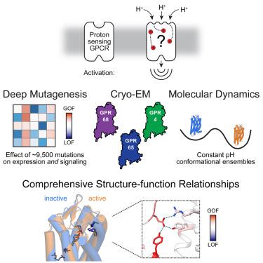
Three proton-sensing G protein-coupled receptors (GPCRs)—GPR4, GPR65, and GPR68—respond to extracellular pH to regulate diverse physiology. How protons activate these receptors is poorly understood. We determined cryogenic-electron microscopy (cryo-EM) structures of each receptor to understand the spatial arrangement of proton-sensing residues. Using deep mutational scanning (DMS), we determined the functional importance of every residue in GPR68 activation by generating ∼9,500 mutants and measuring their effects on signaling and surface expression. Constant-pH molecular dynamics simulations provided insights into the conformational landscape and protonation patterns of key residues. This unbiased approach revealed that, unlike other proton-sensitive channels and receptors, no single site is critical for proton recognition. Instead, a network of titratable residues extends from the extracellular surface to the transmembrane region, converging on canonical motifs to activate proton-sensing GPCRs. Our approach integrating structure, simulations, and unbiased functional interrogation provides a framework for understanding GPCR signaling complexity.
Abstract Image

求助全文
约1分钟内获得全文 求助全文
来源期刊
Cell
Cell 生物-生化与分子生物学
CiteScore
110.00
自引率
0.80%
发文量
396
审稿时长
2 months
期刊介绍: Cells is an international, peer-reviewed, open access journal that focuses on cell biology, molecular biology, and biophysics. It is affiliated with several societies, including the Spanish Society for Biochemistry and Molecular Biology (SEBBM), Nordic Autophagy Society (NAS), Spanish Society of Hematology and Hemotherapy (SEHH), and Society for Regenerative Medicine (Russian Federation) (RPO). The journal publishes research findings of significant importance in various areas of experimental biology, such as cell biology, molecular biology, neuroscience, immunology, virology, microbiology, cancer, human genetics, systems biology, signaling, and disease mechanisms and therapeutics. The primary criterion for considering papers is whether the results contribute to significant conceptual advances or raise thought-provoking questions and hypotheses related to interesting and important biological inquiries. In addition to primary research articles presented in four formats, Cells also features review and opinion articles in its "leading edge" section, discussing recent research advancements and topics of interest to its wide readership.
×
引用
GB/T 7714-2015
复制
MLA
复制
APA
复制
导出至
BibTeX EndNote RefMan NoteFirst NoteExpress
×
提示
您的信息不完整,为了账户安全,请先补充。
现在去补充
×
提示
您因"违规操作"
具体请查看互助需知
我知道了
×
提示
确定
请完成安全验证×
copy
已复制链接
快去分享给好友吧!
我知道了
右上角分享
点击右上角分享
0
联系我们:info@booksci.cn Book学术提供免费学术资源搜索服务,方便国内外学者检索中英文文献。致力于提供最便捷和优质的服务体验。 Copyright © 2023 布克学术 All rights reserved.
京ICP备2023020795号-1
ghs 京公网安备 11010802042870号
Book学术文献互助
Book学术文献互助群
群 号:481959085
Book学术官方微信