{"title":"A Combination of Cell Findings Improves the Accuracy of Voided Urine Cytology.","authors":"Sachiko Iwai, Mitsuru Kinjo","doi":"10.4103/joc.joc_10_24","DOIUrl":null,"url":null,"abstract":"<p><strong>Introduction: </strong>Urine cytology is a morphological diagnostic test that is, patient-friendly and easy to sample but subjective in morphological evaluation. This study aims to evaluate the effect of combining cell findings to assess urine cytology.</p><p><strong>Materials and methods: </strong>Thirty cell findings found in high-grade urothelial carcinoma (HGUC) were selected for morphological abnormalities, each with detailed definitions. All 66 HGUC and 30 benign cases were examined for morphological abnormalities. The number of cases with at least one abnormal cell finding deviating from normal cells was used. The HGUC and benign cases were compared using significance difference tests. Using the cell findings extracted by the exact tests, discriminant analysis was performed to determine the valid cell findings for HGUC diagnosis.</p><p><strong>Results: </strong>There was a difference in the detection rate of abnormal findings between HGUC and benign cases. Additionally, 20 cell findings were detected significantly more frequently in patients with HGUC. Furthermore, the discriminant analysis revealed that six cellular findings (high nucleus-to-cytoplasm ratio, hyperchromasia, eccentric nuclei, nuclear protrusion, unevenly distributed chromatin, and irregular nuclear shape) showed high accuracy in confirming the diagnosis of HGUC.</p><p><strong>Conclusions: </strong>Five- to six-cell findings were individually valid; however, the combination of cell findings is crucial for an objective and accurate diagnosis of HGUC.</p>","PeriodicalId":50217,"journal":{"name":"Journal of Cytology","volume":"41 4","pages":"183-191"},"PeriodicalIF":1.0000,"publicationDate":"2024-10-01","publicationTypes":"Journal Article","fieldsOfStudy":null,"isOpenAccess":false,"openAccessPdf":"https://www.ncbi.nlm.nih.gov/pmc/articles/PMC11676093/pdf/","citationCount":"0","resultStr":null,"platform":"Semanticscholar","paperid":null,"PeriodicalName":"Journal of Cytology","FirstCategoryId":"3","ListUrlMain":"https://doi.org/10.4103/joc.joc_10_24","RegionNum":4,"RegionCategory":"医学","ArticlePicture":[],"TitleCN":null,"AbstractTextCN":null,"PMCID":null,"EPubDate":"2024/11/8 0:00:00","PubModel":"Epub","JCR":"Q4","JCRName":"MEDICAL LABORATORY TECHNOLOGY","Score":null,"Total":0}
引用次数: 0
Abstract
Introduction: Urine cytology is a morphological diagnostic test that is, patient-friendly and easy to sample but subjective in morphological evaluation. This study aims to evaluate the effect of combining cell findings to assess urine cytology.
Materials and methods: Thirty cell findings found in high-grade urothelial carcinoma (HGUC) were selected for morphological abnormalities, each with detailed definitions. All 66 HGUC and 30 benign cases were examined for morphological abnormalities. The number of cases with at least one abnormal cell finding deviating from normal cells was used. The HGUC and benign cases were compared using significance difference tests. Using the cell findings extracted by the exact tests, discriminant analysis was performed to determine the valid cell findings for HGUC diagnosis.
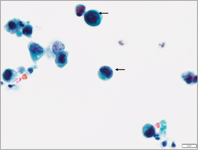
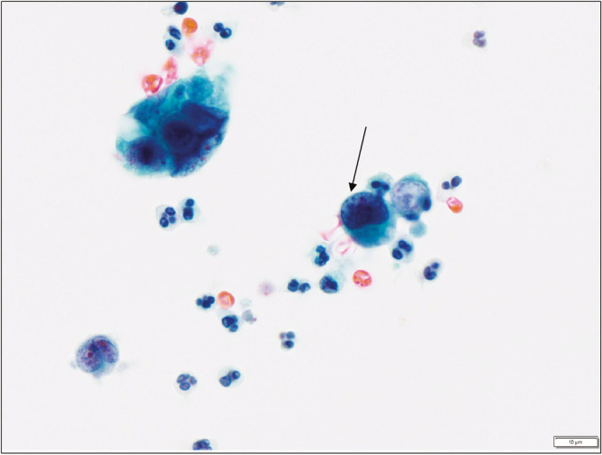
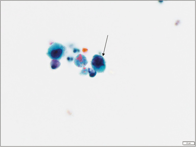
Results: There was a difference in the detection rate of abnormal findings between HGUC and benign cases. Additionally, 20 cell findings were detected significantly more frequently in patients with HGUC. Furthermore, the discriminant analysis revealed that six cellular findings (high nucleus-to-cytoplasm ratio, hyperchromasia, eccentric nuclei, nuclear protrusion, unevenly distributed chromatin, and irregular nuclear shape) showed high accuracy in confirming the diagnosis of HGUC.
Conclusions: Five- to six-cell findings were individually valid; however, the combination of cell findings is crucial for an objective and accurate diagnosis of HGUC.
期刊介绍:
The Journal of Cytology is the official Quarterly publication of the Indian Academy of Cytologists. It is in the 25th year of publication in the year 2008. The journal covers all aspects of diagnostic cytology, including fine needle aspiration cytology, gynecological and non-gynecological cytology. Articles on ancillary techniques, like cytochemistry, immunocytochemistry, electron microscopy, molecular cytopathology, as applied to cytological material are also welcome. The journal gives preference to clinically oriented studies over experimental and animal studies. The Journal would publish peer-reviewed original research papers, case reports, systematic reviews, meta-analysis, and debates.
 求助内容:
求助内容: 应助结果提醒方式:


