Fluorescence Interference-Based Polarized Structured Illumination Microscopy for High Axial Accuracy Morphology Imaging of Dipole Orientations
IF 6.5
1区 物理与天体物理
Q1 MATERIALS SCIENCE, MULTIDISCIPLINARY
引用次数: 0
Abstract
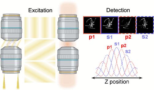
The dipole orientation of fluorescent molecules reveals the structural organization at the subcellular level, and imaging its distribution is important for fundamental cell biology studies. Using conventional fluorescence microscopy, the orientation of fluorescent dipoles can be obtained by demodulating the polarization states through excitation or emission processes. The recent introduction of structured illumination microscopy (SIM) has improved the axial and lateral resolution to around 300 and 100 nm, respectively. However, the axial resolution is still very limited, which hinders the capabilities to offer more precise measurements of polarization information. Here, we report Fluorescence Interference based polarized structured illumination microscopy (FI-pSIM), which provides a sub-30 nm axial reconstruction accuracy and maintains the same lateral resolution comparable to SIM by only 9 raw images. By synergizing the fluorescence interference via the 4Pi configuration and the polarization demodulation of structured illumination, FI-pSIM enables three-dimensional morphological imaging of ensemble dipole orientations in subcellular structures with ∼ sub-30 nm accuracy of axial reconstruction. The high efficacy and four-dimensional (4D) imaging modality of FI-pSIM are demonstrated by mapping the distribution and orientation of fluorescent dipoles in biological microfilaments. By elucidating the dipole orientations, we envision FI-pSIM may open up new avenues for demystifying the 4D organizations of subcellular structures.
基于荧光干涉的极化结构照明显微镜用于偶极子取向的高轴向精度形态学成像
荧光分子的偶极取向揭示了亚细胞水平的结构组织,其分布成像对细胞生物学的基础研究具有重要意义。利用传统的荧光显微镜,可以通过激发或发射过程解调偏振态来获得荧光偶极子的取向。最近引入的结构照明显微镜(SIM)将轴向和横向分辨率分别提高到300 nm和100 nm左右。然而,轴向分辨率仍然非常有限,这阻碍了提供更精确的偏振信息测量的能力。在这里,我们报道了基于荧光干涉的偏振结构照明显微镜(FI-pSIM),它提供了低于30 nm的轴向重建精度,并且仅通过9张原始图像就保持了与SIM相同的横向分辨率。通过4Pi结构的荧光干涉和结构照明的偏振解调的协同作用,FI-pSIM实现了亚细胞结构中集合偶极子取向的三维形态成像,轴向重建精度为~ sub- 30nm。通过绘制生物微丝中荧光偶极子的分布和取向,证明了FI-pSIM的高效率和四维成像模式。通过阐明偶极子取向,我们设想FI-pSIM可能为揭示亚细胞结构的4D组织开辟新的途径。
本文章由计算机程序翻译,如有差异,请以英文原文为准。
求助全文
约1分钟内获得全文
求助全文
来源期刊
ACS Photonics
NANOSCIENCE & NANOTECHNOLOGY-MATERIALS SCIENCE, MULTIDISCIPLINARY
CiteScore
11.90
自引率
5.70%
发文量
438
审稿时长
2.3 months
期刊介绍:
Published as soon as accepted and summarized in monthly issues, ACS Photonics will publish Research Articles, Letters, Perspectives, and Reviews, to encompass the full scope of published research in this field.
 求助内容:
求助内容: 应助结果提醒方式:


