A Rare Case of a Malignant Proliferating Trichilemmal Tumor: A Molecular Study Harboring Potential Therapeutic Significance and a Review of Literature.
Mokhtar H Abdelhammed, Hanna Siatecka, A Hafeez Diwan, Christie J Finch, Angela D Haskins, David J Hernandez, Ya Xu
{"title":"A Rare Case of a Malignant Proliferating Trichilemmal Tumor: A Molecular Study Harboring Potential Therapeutic Significance and a Review of Literature.","authors":"Mokhtar H Abdelhammed, Hanna Siatecka, A Hafeez Diwan, Christie J Finch, Angela D Haskins, David J Hernandez, Ya Xu","doi":"10.3390/dermatopathology11040038","DOIUrl":null,"url":null,"abstract":"<p><p>Malignant proliferating trichilemmal tumors (MPTTs), arising from the external root sheath of hair follicles, are exceptionally rare, with limited documentation of their genetic alterations. We present a case of a 64-year-old African American woman who initially presented with a gradually enlarging nodule on her posterior scalp. An initial biopsy at an outside hospital suggested metastatic adenocarcinoma or squamous cell carcinoma (SCC) of an uncertain origin. A subsequent wide local excision revealed a 2.0 cm tumor demonstrating characteristic trichilemmal keratinization, characterized by an abrupt transition from the nucleated epithelium to a laminated keratinized layer, confirming MPTT. Immunohistochemistry demonstrated diffuse p53 expression, patchy CD 34 expression, focal HER2 membranous expression, and patchy p16 staining (negative HPV ISH). A molecular analysis identified TP53 mutation and amplifications in the ERBB2 (HER2), BRD4, and TYMS. Additional gene mutations of uncertain significance included HSPH1, ATM, PDCD1 (PD-1), BARD1, MSH3, LRP1B, KMT2C (MLL3), GNA11, and RUNX1. Assessments for the homologous recombination deficiency, PD-L1 expression, gene rearrangement, altered splicing, and DNA mismatch repair gene expression were negative. The confirmation of ERBB2 (HER2) amplification in the MPTT through a molecular analysis suggests potential therapeutic avenues involving anti-HER2 monoclonal antibodies. The presence of the TP53 mutation, without the concurrent gene mutations typically observed in SCC, significantly aided in this differential diagnosis.</p>","PeriodicalId":42885,"journal":{"name":"Dermatopathology","volume":"11 4","pages":"354-363"},"PeriodicalIF":1.7000,"publicationDate":"2024-12-10","publicationTypes":"Journal Article","fieldsOfStudy":null,"isOpenAccess":false,"openAccessPdf":"https://www.ncbi.nlm.nih.gov/pmc/articles/PMC11674666/pdf/","citationCount":"0","resultStr":null,"platform":"Semanticscholar","paperid":null,"PeriodicalName":"Dermatopathology","FirstCategoryId":"1085","ListUrlMain":"https://doi.org/10.3390/dermatopathology11040038","RegionNum":0,"RegionCategory":null,"ArticlePicture":[],"TitleCN":null,"AbstractTextCN":null,"PMCID":null,"EPubDate":"","PubModel":"","JCR":"Q3","JCRName":"DERMATOLOGY","Score":null,"Total":0}
引用次数: 0
Abstract
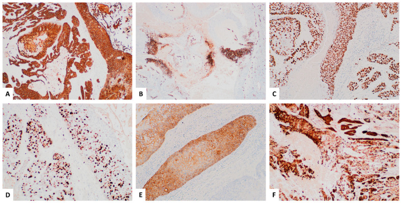
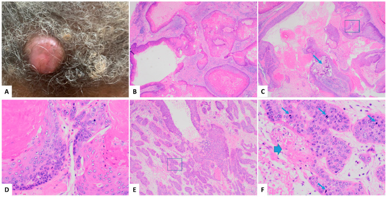
Malignant proliferating trichilemmal tumors (MPTTs), arising from the external root sheath of hair follicles, are exceptionally rare, with limited documentation of their genetic alterations. We present a case of a 64-year-old African American woman who initially presented with a gradually enlarging nodule on her posterior scalp. An initial biopsy at an outside hospital suggested metastatic adenocarcinoma or squamous cell carcinoma (SCC) of an uncertain origin. A subsequent wide local excision revealed a 2.0 cm tumor demonstrating characteristic trichilemmal keratinization, characterized by an abrupt transition from the nucleated epithelium to a laminated keratinized layer, confirming MPTT. Immunohistochemistry demonstrated diffuse p53 expression, patchy CD 34 expression, focal HER2 membranous expression, and patchy p16 staining (negative HPV ISH). A molecular analysis identified TP53 mutation and amplifications in the ERBB2 (HER2), BRD4, and TYMS. Additional gene mutations of uncertain significance included HSPH1, ATM, PDCD1 (PD-1), BARD1, MSH3, LRP1B, KMT2C (MLL3), GNA11, and RUNX1. Assessments for the homologous recombination deficiency, PD-L1 expression, gene rearrangement, altered splicing, and DNA mismatch repair gene expression were negative. The confirmation of ERBB2 (HER2) amplification in the MPTT through a molecular analysis suggests potential therapeutic avenues involving anti-HER2 monoclonal antibodies. The presence of the TP53 mutation, without the concurrent gene mutations typically observed in SCC, significantly aided in this differential diagnosis.

 求助内容:
求助内容: 应助结果提醒方式:


