{"title":"Decoding Brain Development and Aging: Pioneering Insights From MRI Techniques.","authors":"Akifumi Hagiwara, Satoru Kamio, Junko Kikuta, Moto Nakaya, Wataru Uchida, Shohei Fujita, Stikov Nikola, Toshiaki Akasahi, Akihiko Wada, Koji Kamagata, Shigeki Aoki","doi":"10.1097/RLI.0000000000001120","DOIUrl":null,"url":null,"abstract":"<p><strong>Abstract: </strong>The aging process induces a variety of changes in the brain detectable by magnetic resonance imaging (MRI). These changes include alterations in brain volume, fluid-attenuated inversion recovery (FLAIR) white matter hyperintense lesions, and variations in tissue properties such as relaxivity, myelin, iron content, neurite density, and other microstructures. Each MRI technique offers unique insights into the structural and compositional changes occurring in the brain due to normal aging or neurodegenerative diseases. Age-related brain volume changes encompass a decrease in gray matter and an increase in ventricular volume, associated with cognitive decline. White matter hyperintensities, detected by FLAIR, are common and linked to cognitive impairments and increased risk of stroke and dementia. Tissue relaxometry reveals age-related changes in relaxivity, aiding the distinction between normal aging and pathological conditions. Myelin content, measurable by MRI, changes with age and is associated with cognitive and motor function alterations. Iron accumulation, detected by susceptibility-sensitive MRI, increases in certain brain regions with age, potentially contributing to neurodegenerative processes. Diffusion MRI provides detailed insights into microstructural changes such as neurite density and orientation. Neurofluid imaging, using techniques like gadolinium-based contrast agents and diffusion MRI, reveals age-related changes in cerebrospinal and interstitial fluid dynamics, crucial for brain health and waste clearance. This review offers a comprehensive overview of age-related brain changes revealed by various MRI techniques. Understanding these changes helps differentiate between normal aging and pathological conditions, aiding the development of interventions to mitigate age-related cognitive decline and other symptoms. Recent advances in machine learning and artificial intelligence have enabled novel methods for estimating brain age, offering also potential biomarkers for neurological and psychiatric disorders.</p>","PeriodicalId":14486,"journal":{"name":"Investigative Radiology","volume":" ","pages":"162-174"},"PeriodicalIF":8.0000,"publicationDate":"2025-03-01","publicationTypes":"Journal Article","fieldsOfStudy":null,"isOpenAccess":false,"openAccessPdf":"https://www.ncbi.nlm.nih.gov/pmc/articles/PMC11801466/pdf/","citationCount":"0","resultStr":null,"platform":"Semanticscholar","paperid":null,"PeriodicalName":"Investigative Radiology","FirstCategoryId":"3","ListUrlMain":"https://doi.org/10.1097/RLI.0000000000001120","RegionNum":1,"RegionCategory":"医学","ArticlePicture":[],"TitleCN":null,"AbstractTextCN":null,"PMCID":null,"EPubDate":"2024/10/9 0:00:00","PubModel":"Epub","JCR":"Q1","JCRName":"RADIOLOGY, NUCLEAR MEDICINE & MEDICAL IMAGING","Score":null,"Total":0}
引用次数: 0
Abstract
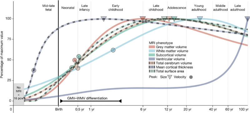
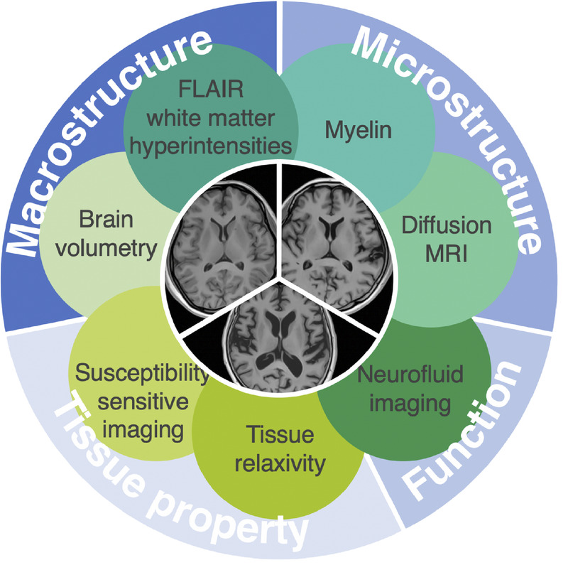
Abstract: The aging process induces a variety of changes in the brain detectable by magnetic resonance imaging (MRI). These changes include alterations in brain volume, fluid-attenuated inversion recovery (FLAIR) white matter hyperintense lesions, and variations in tissue properties such as relaxivity, myelin, iron content, neurite density, and other microstructures. Each MRI technique offers unique insights into the structural and compositional changes occurring in the brain due to normal aging or neurodegenerative diseases. Age-related brain volume changes encompass a decrease in gray matter and an increase in ventricular volume, associated with cognitive decline. White matter hyperintensities, detected by FLAIR, are common and linked to cognitive impairments and increased risk of stroke and dementia. Tissue relaxometry reveals age-related changes in relaxivity, aiding the distinction between normal aging and pathological conditions. Myelin content, measurable by MRI, changes with age and is associated with cognitive and motor function alterations. Iron accumulation, detected by susceptibility-sensitive MRI, increases in certain brain regions with age, potentially contributing to neurodegenerative processes. Diffusion MRI provides detailed insights into microstructural changes such as neurite density and orientation. Neurofluid imaging, using techniques like gadolinium-based contrast agents and diffusion MRI, reveals age-related changes in cerebrospinal and interstitial fluid dynamics, crucial for brain health and waste clearance. This review offers a comprehensive overview of age-related brain changes revealed by various MRI techniques. Understanding these changes helps differentiate between normal aging and pathological conditions, aiding the development of interventions to mitigate age-related cognitive decline and other symptoms. Recent advances in machine learning and artificial intelligence have enabled novel methods for estimating brain age, offering also potential biomarkers for neurological and psychiatric disorders.
期刊介绍:
Investigative Radiology publishes original, peer-reviewed reports on clinical and laboratory investigations in diagnostic imaging, the diagnostic use of radioactive isotopes, computed tomography, positron emission tomography, magnetic resonance imaging, ultrasound, digital subtraction angiography, and related modalities. Emphasis is on early and timely publication. Primarily research-oriented, the journal also includes a wide variety of features of interest to clinical radiologists.

 求助内容:
求助内容: 应助结果提醒方式:


